Biological Activity
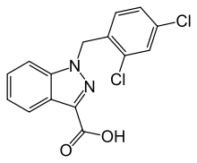
Lonidamine (AF-1890) is a novel CFTR open channel blocker with an IC50 of 0.85 mM, which inhibits aerobic glycolysis in cancer cells. Lonidamine (AF-1890) is a hexokinase and mitochondrial pyruvate carrier inhibitor (Ki=2.5 μM). Lonidamine could reduce the dose of temozolomide required for radiosensitization of brain tumours. Lonidamine elicits ERK and Akt/mTOR pathway activation, as indicated by increased ERK, Akt, p70S6K and rpS6 phosphorylation, and these effects are reduced by co-treatment with the anti-leukemic agent arsenic trioxide. Lonidamine significantly extends both median and maximum lifespan of C. elegans when applied at a concentration of 5 micromolar by 8% each. Lonidamine, a dechlorinate derivative of indazole-3-carboxylic acid, has proved to exert a powerful antiproliferative effect and to impair the energy metabolism of neoplastic cells.
Customer Product Validations & Biological Datas
. figure 14.jpg) |
Source |
Uinversity of Bergen thesis (2016). Figure 14.Lonidamine (AbMole BioScience, CAS No.: 50264-69-2) |
Method |
WST-1 assay |
Cell Lines |
HL60, MOLM13, NB4, and PBMC cells |
Concentrations |
1~100 µM |
Incubation Time |
48 h |
Results |
An overall higher degree of reduced metabolic activity was observed from three independent studies with MOLM13 (Figure 14A) and NB4 (Figure14B) cells thus were suggested being more responsive to co-treatment. HL60 cells responded revealed low response to both drugs alone,resulting in only 10-25 % reduced proliferation when exposed to the highest concentrations of VPA (1 mM) and LND (100 µM), respectively (Figure 14C). |
. figure 13.jpg) |
Source |
Uinversity of Bergen thesis (2016). Figure 13.Lonidamine (AbMole BioScience, CAS No.: 50264-69-2) |
Method |
Cell viability assay |
Cell Lines |
HL60, MOLM13, NB4, and PBMC cells |
Concentrations |
1~100 µM |
Incubation Time |
48 h |
Results |
These data indicated a comparable trend of viability in all three cell lines following combinatorial treatment of VPA and LND (Figure 13B, C). HL60 cells are only presented for Hoechst viability scoring in figure 13A since the cells do not expose phosphatidylserine upon apoptosis induction. |
. figure 12.jpg) |
Source |
Uinversity of Bergen thesis (2016). Figure 12.Lonidamine (AbMole BioScience, CAS No.: 50264-69-2) |
Method |
MST-1 proliferation assay |
Cell Lines |
HL60 cells |
Concentrations |
50 and 100 µM |
Incubation Time |
48 or 72 h |
Results |
Maximal potentiation of LND was obtained by 100 µM LND in the sequence VPA LND+VPA, but no significant increased anti-proliferative effect was obtained from three independent and replicable studies. |
. figure 11.jpg) |
Source |
Uinversity of Bergen thesis (2016). Figure 11.Lonidamine (AbMole BioScience, CAS No.: 50264-69-2) |
Method |
Hoechst 33342 staining |
Cell Lines |
HL60, MOLM13 and NB5 cells |
Concentrations |
0~500 µM |
Incubation Time |
24 or 48 h |
Results |
LND elicits potential of inducing apoptosis after both 24 hours and 48 hour incubation at higher concentrations (> 50 µM) in HL60, MOLM13 and NB4 (Figure 11A-B). Apoptotic cells in the stage of morphological changes such as fragmented or condensed nuclei are shown in Figure 11C, visualized by fluorescence microscopy. |
. figure 10.jpg) |
Source |
Uinversity of Bergen thesis (2016). Figure 10.Lonidamine (AbMole BioScience, CAS No.: 50264-69-2) |
Method |
WST-1 proliferation assay |
Cell Lines |
HL60, MOLM13, NB4, and PBMC cells |
Concentrations |
0~1000 µM |
Incubation Time |
48 h |
Results |
The distinct response of PBMC comparable to MOLM13 and HL60 is noticeably demonstrated in Figure 10A. NB4 was significantly (P<0.05) more sensitive to LND under 24 hours of exposure than 48 hours (5 – 500 µM). The IC50 of LND in NB4 (201 µM), HL60 (248 µM) and MOLM13 (124 µM) are presented in Figure 10 B, C and D indicating LND to be more effective after 48 hour treatment in MOLM13 and NB4 cells at lower concentrations than when treated for 24 hours. |
. figure 9.jpg) |
Source |
Uinversity of Bergen thesis (2016). Figure 9.Lonidamine (AbMole BioScience, CAS No.: 50264-69-2) |
Method |
WST-1 proliferation assay |
Cell Lines |
HL60, MOLM13, NB4, and PBMC cells |
Concentrations |
0~1000 µM |
Incubation Time |
24 h |
Results |
No significant difference (P>0.05) in reduced metabolically active cells was obtained between HL60 and NB4 cells after 24 hour treatment with LND. IC50s of LND was obtained from WST-1 proliferation assay, and are presented in Figure 9B, C and D for HL60 (149 µM), MOLM13 (218 µM) and NB4 (203 µM), respectively. A wider 95% CI is observed for the IC50 of LND in treated NB4 cells (Figure 9D) than for HL60 and MOLM13, thus we cannot exclude the possibility that the IC50 of NB4 could be present anywhere between 106 – 387 µM. |
. figure 7.jpg) |
Source |
Uinversity of Bergen thesis (2016). Figure 7.Lonidamine (AbMole BioScience, CAS No.: 50264-69-2) |
Method |
WST-1 assay |
Cell Lines |
NB4 cells |
Concentrations |
12.5, 25, 50, 75 and 100 µM |
Incubation Time |
48 h |
Results |
The combined effect of HU and MMF, shown in Figure 7A, followed the same anti-proliferative response to that of MMF alone and no additive effect or synergism was achieved. As both HK1 and HPRT1 were down-regulated by VPA in BNML rats, the combination of LND and MMF (Figure 7B) was investigated for potential synergistic or additive effect on proliferation and apoptosis. |
Chemical Information
Molecular Weight |
321.16 |
Formula |
C15H10Cl2N2O2 |
CAS Number |
50264-69-2
|
Solubility (25°C) |
DMSO 44 mg/mL |
Storage |
Powder -20°C 3 years ; 4°C 2 years
In solvent -80°C 6 months ; -20°C 1 month
|
References
[1] Calviño E, et al. Biochem Pharmacol. Increased apoptotic efficacy of lonidamine plus arsenic trioxide combination in human leukemia cells. Reactive oxygen species generation and defensive protein kinase (MEK/ERK, Akt/mTOR) modulation.
[2] Di Cosimo S, et al. Drugs Today (Barc). Lonidamine: efficacy and safety in clinical trials for the treatment of solid tumors.
[3] Gong X, et al. Br J Pharmacol. Mechanism of lonidamine inhibition of the CFTR chloride channel.