Free shipping on all orders over $ 500

Calcein Red, AM

Cat. No. M10356

All AbMole products are for research use only, cannot be used for human consumption.

Calcein Red, AM Structure
Size Price Availability Quantity
1mg USD 550  USD550 In stock
Free Delivery on orders over USD 500 Bulk Inquiry?

Quality Control & Documentation
  • Purity: Biological Stain
  • COA
  • MSDS
Biological Activity

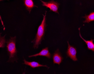
Calcein AM is one of the most popular fluorescent probes used for labeling and monitoring cellular functions of live cells. Non-fluorescent Calcein Red AM can easily get into live cells and hydrolyzes to generate strongly fluorescent Calcein Red dye. Calcein Red dye can be monitored with the common TRITC/Cy3 filter set.


PREPARATION OF STOCK SOLUTIONS

Unless otherwise noted, all unused stock solutions should be divided into single-use aliquots and stored at -20 °C after preparation. Avoid repeated freeze-thaw cycles.
Calcein Red AM Stock Solution
Prepare a 2 to 5 mM stock solution of Calcein Red AM in high-quality, anhydrous DMSO.
Note The nonionic detergent Pluronic® F-127 can be used to increase the aqueous solubility of AM esters. In the staining buffer, the final Pluronic® F-127 concentration should be approximately 0.02%. A variety of Pluronic® F-127 products can be purchased from AAT Bioquest. Avoid long-term storage of AM esters in the presence of Pluronic® F-127.

PREPARATION OF WORKING SOLUTION

Calcein Red AM Working Solution
Prepare a Calcein Red AM working solution of 1 to 10 µM in the buffer of your choice (e.g., Hanks and Hepes buffer). For most cell lines, Calcein Red AM at the final concentration of 4 to 5 µM is recommended. The exact concentration of indicators required for cell loading must be determined empirically.
Note If your cells contain organic anion-transporters, probenecid (1–2.5 mM) or sulfinpyrazone (0.1–0.25 mM) may be added to the working solution to reduce leakage of the de-esterified indicators.

SAMPLE EXPERIMENTAL PROTOCOL

  1. Prepare cells for imaging.
  2. Remove the cell culture medium and wash cells once with serum-free buffer to remove any remaining media.
    Note Serum in cell culture media may contain esterase activity, which can increase background interference.
  3. Add Calcein Red AM working solution to the culture.
  4. Incubate cells at 37 °C for 30 to 60 minutes.
  5. Replace the dye working solution with HHBS or buffer of your choice (containing an anion transporter inhibitor, such as 1 mM probenecid, if applicable) to remove any excess probes.
  6. Measure the fluorescence intensity using either a fluorescence microscope equipped with a TRITC filter set, a flow cytometer equipped with green/yellow laser and a 585/40 nm filter, or a fluorescence plate reader at Ex/Em = 540/590 nm cutoff 570 nm.

EXAMPLE DATA ANALYSIS AND FIGURES


Figure 1. Images of Live HeLa cells stained with Calcein Red, AM (Cat# M10356). Cell nuclei were stained with Hoechst 33342 (Blue, Cat# M5112).


Chemical Information
Molecular Weight 1015.79
Solubility (25°C) DMSO 1~5mM
Storage -20°C, protect from light, dry, sealed
References

[1] Nienke R Wevers, et al. Fluids Barriers CNS. A perfused human blood-brain barrier on-a-chip for high-throughput assessment of barrier function and antibody transport

[2] Hongli Li, et al. Toxicol Appl Pharmacol. Astragaloside IV protects blood-brain barrier integrity from LPS-induced disruption via activating Nrf2 antioxidant signaling pathway in mice

Related Fluorescent Dye Products
4-methylumbelliferyl α-D-glucopyranoside

4-Methylumbelliferyl-α-D-Glucopyranoside is a fluorescent substrate for α-glucosidase (GAA), which releases the fluorescent moiety 4-methylumbelliferyl (4-MU) upon cleavage. 4-MU has pH-dependent fluorescence excitation activity, with excitation wavelengths of 320 nm at low pH (1.97-6.72) and 360 nm at high pH (7.12-10.3), respectively.

Cyanine7 amine

Cyanine7 amine is a near infrared dye with free amine group for the conjμgation with activated esters and other reactive molecules.

BODIPY TR Cadaverine

BODIPY TR Cadaverine is a red fluorescent dye. BODIPY TR Cadaverine can be used in a a highly sensitive and robust fluorescent displacement assay, which binds to native LPS strongly, specifically recognizing lipid A.

Sudan Black B

Sudan Black B, a fat-soluble diazo dye, is a histochemical stain. Sudan Black B can be used for staining of neutral triglycerides and lipids.

DASPEI

DASPEI is a cationic styrenyl mitochondrial dye. DASPEI has excitation and emission wavelength at 550/573 nm, which has good light chromogenic property. DASPEI can stain mitochondria in living cells with good labeling property. DASPEI can also be used to stain presynaptic nerve endings independently of neuronal activity.

  Catalog
Abmole Inhibitor Catalog


Keywords: Calcein Red, AM supplier, Fluorescent Dye, inhibitors, activators

All AbMole products are for research use only, cannot be used for human consumption or veterinary use. We do not provide products or services to individuals. Please comply with the intended use and do not use AbMole products for any other purpose.


Products are for research use only. Not for human use. We do not sell to patients.
© Copyright 2010-2024 AbMole BioScience. All Rights Reserved.