All AbMole products are for research use only, cannot be used for human consumption.
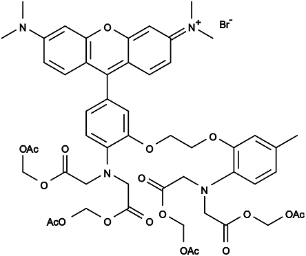
Rhod-2 AM is a fluorescent, mitochondrial probe (λex=552 nm, λem=581 nm). Rhod-2 AM is cell-permeable version of Rhod-2. The long-wavelength Rhod-2 Ca2+ indicators are valuable alternatives to Fluo-3 for experiments in cells and tissues that have high levels of autofluorescence. Fluorescent probes that show spectral responses upon binding Ca2+ have enabled researchers to investigate changes in intracellular free Ca2+ concentrations by using fluorescence microscopy, flow cytometry, fluorescence spectroscopy and fluorescence microplate readers.

Exp Neurol. 2024 May 3.
Mfn2 regulates mitochondria and mitochondria-associated endoplasmic reticulum membrane function in neurodegeneration induced by repeated sevoflurane exposure
Rhod-2 AM purchased from AbMole
| Molecular Weight | 1123.96 |
| Formula | C52H59BrN4O |
| CAS Number | 145037-81-6 |
| Solubility (25°C) | DMSO 1~5mM |
| Storage | -20°C, protect from light, dry, sealed |
| Related Fluorescent Dye Products |
|---|
| pH Fluorescent Probe Red 600, SE
pH Fluorescent Probe Red 600, SE is a pH-sensitive fluorescent dye, the fluorescence intensity changes significantly with changes in the pH of the environment. pH Fluorescent Probe Red 600, SE is weakly fluorescent outside the cells, but its fluorescence is significantly enhanced in acidic compartments (such as phagosomes, lysosomes and endosomes). It can be used for multiplexing cellular functional analysis with green dyes such as GFP, Fluo-8, calcein, or FITC-labeled antibodies. Ex (nm) 576, Em (nm) 597 |
| DSPE-Rhodamine
DSPE-Rhodamine is formed by the conjugation of DSPE with the Rhodamine fluorescent dye. DSPE possesses a hydrophobic lipid tail and a hydrophilic head group, while Rhodamine is a red fluorescent dye. DSPE-Rhodamine retains the lipid properties of DSPE while also imparting fluorescent labelling capabilities. As a key component of delivery systems, DSPE-Rhodamine enhances targeting and bioavailability. Its lipid properties facilitate the penetration of molecules through cell membranes, while its fluorescent properties enable tracking of distribution and dynamic changes within the body. |
| LD540
LD540 is a novel high-sensitivity lipophilic dye modified with BODIPY fluorescent groups, designed for precise labelling and imaging of lipid droplets. LD540 exhibits excellent photostability and an optimal fluorescence spectrum, making it compatible with various commonly used fluorescent dyes (such as DAPI and Alexa Fluor 647), thereby supporting multi-colour imaging requirements. Additionally, LD540 is suitable for both fixed and live cells and can label ultra-small lipid droplets. |
| PDMPO
PDMPO (Yellow/Blue DND-160) is a ratiometric probe for the determination of lysosomal pH for fluorescence imaging. PDMPO exhibits pH-dependent dual excitation and dual emission peaks. PDMPO produces blue fluorescence (Ex/Em=329 nm/440 nm) in weakly acidic organelles, and in more acidic lysosomes it becomes yellow fluorescence (Ex/Em=384 nm/540 nm). |
| Copper probe CF4
Copper probe CF4 (Copper fluor CF4) is a Cu+-specific fluorescent probe based on a rhodol dye scaffold. Copper probe CF4 (Copper fluor CF4) has high copper selectivity with a Kd value of 2.9×10⁻¹³ M, particularly over zinc and iron, as well as abundant cellular alkali and alkaline earth metals. Copper probe CF4 (Copper fluor CF4) is stable in a physiologically relevant pH regime between 6 and 8 (wavelengths of 415 nm for excitation and 660 nm for emission). Copper probe CF4 (Copper fluor CF4) can be used to study colon cancer. |

All AbMole products are for research use only, cannot be used for human consumption or veterinary use. We do not provide products or services to individuals. Please comply with the intended use and do not use AbMole products for any other purpose.


Products are for research use only. Not for human use. We do not sell to patients.
© Copyright 2010-2024 AbMole BioScience. All Rights Reserved.