Intended Use
Cell Counting Kit-8 (CCK-8) allows sensitive colorimetric assays for the determination of cell viability in cell proliferation and cytotoxicity assays.
General Description
CCK solution is a one-bottle solution; no pre-mixing of components is required. CCK, being nonradioactive, allows sensitive colorimetric assays for the determination of the number of viable cells in cell proliferation and cytotoxicity assays. WSTS* is reduced by dehydrogenases in cells to give an orange colored product (formazan), which is soluble in tissue culture medium. The amount of formazan dye generated by dehydrogenases in cells is directly proportional to the number of living cells.
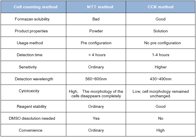
Comparison between CCK method and MTT method:
Protocol:
1. Usually in the 96 hole plate, cell proliferation and cytotoxicity test are inoculated with cell suspension (5000cells and 2000 cells /100 uL/ hole, the specific number of the cells per hole should be decided according to the size and speed of the cell proliferation speed and other factors). The system is filled with a corresponding amount of cell culture medium, drug and CCK-8 solution, and the holes without cells are regarded as a blank control.
2. The cells adhered to the wall for 24 hours and are given a specific dose of 0-10 mg of stimulation in accordance with the experimental requirements.
3. The drugs are treated for 2~4 days and then add 10 uL of CCK solution at each hole (be careful not to generate bubbles in the pores, which will affect OD value)
4. Incubate the incubator for 1~4 hours, then use the microplate reader to determine the absorbance at 450nm.
5. If you do not detect OD value
temporarily, you may add 10 μL 0.1M HCL solution or 1% w/v SDS
solution, cover it with culture plate and keep it from light at room
temperature. Absorbance does not change in 24 hours.
6. The absorbance of phenol red can be eliminated by blank hole so that the result will not be affected.
Announcements:
1. When using 96 hole plate to detect , if the cell culture time is longer,we must pay attention to evaporation problems. On the one hand, a circle surrounding 96 hole plate is more likely to evaporate, you can take it out, adding the same amount of PBS, water or liquid culture instead; On the other hand, the 96 hole plate can be placed near the incubator water to reduce evaporation.
2. The detection of this kit depends on the reaction catalyzed by dehydrogenase, so reducing agents (such as some antioxidants) will interfere with the detection.
3. If there are many reducing agents in the detection system, we should try to remove them.
4. Before using Microplate Reader, you should make sure there is no bubble in each hole, otherwise it will interfere with the determination.
5. This product is limited to scientific research of professionals and should not be used for clinical diagnosis and treatment or used as food and medicine, it should not be deposited in ordinary houses.
6. For your safety and health, please wear lab-gown and disposable gloves.
Vitality calculation:
Cell viability * (%) =[A (dosing) -A (blank)]/[A (0 dosing) -A (blank) * 100%
A (dosing): Absorbance of cells, CCK solutions, and drug solutions
A (blank): Absorbance of the culture medium and CCK solution without cells
A (0 dosing): Absorbance of cells, CCK solutions without drug solutions
* Cell viability: Cell proliferation activity or cytotoxicity activity
Storage conditions:
4 degrees centigrades for one year; -20 degrees centigrades, protected from light for two years.
Documentation Download

Adv Sci (Weinh). 2025 Feb 03.
Targeting FDFT1 Reduces Cholesterol and Bile Acid Production and Delays Hepatocellular Carcinoma Progression Through the HNF4A/ALDOB/AKT1 Axis
Cell Counting Kit-8 purchased from AbMole

Adv Sci (Weinh). 2025 Jun 05; .
Copper‐Based Nanotubes That Enhance Starvation Therapy Through Cuproptosis for Synergistic Cancer Treatment
Cell Counting Kit-8 purchased from AbMole

Acta Biomater. 2025 Jan 26.
Lung-targeted delivery of PTEN mRNA combined with anti-PD-1-mediated immunotherapy for In Situ lung cancer treatmen
Cell Counting Kit-8 purchased from AbMole

Acta Biomater. 2025 Mar 15; .
Immuno-Engineered Macrophage Membrane-Coated Nanodrug to Restore Immune Balance for Rheumatoid Arthritis Treatment
Cell Counting Kit-8 purchased from AbMole

Bioresour Technol. 2025 Jun 16; .
Spirulina subsalsa polysaccharide: self-assembling hydrogel material for immunotherapy applications
Cell Counting Kit-8 purchased from AbMole

Int J Biol Macromol. 2025 Jun 27; .
Acteoside suppresses hepatocellular carcinoma progression via modulation of macrophage migration inhibitory factor and mitogen-activated protein kinase proteins
Cell Counting Kit-8 purchased from AbMole

Cell Death Dis. 2025 Apr 07;16(1):261.
SIRT5-mediated BCAT1 desuccinylation and stabilization leads to ferroptosis insensitivity and promotes cell proliferation in glioma
Cell Counting Kit-8 purchased from AbMole

Oncogene. 2025 Mar 03; .
RBM39 promotes hepatocarcinogenesis by regulating RFX1’s alternative splicing and subsequent activation of integrin signaling pathway
Cell Counting Kit-8 purchased from AbMole

NPJ Precis Oncol. 2025 May 09;9(1):135.
SHMT inhibitor synergizes with 5-Fu to suppress gastric cancer via cell cycle arrest and chemoresistance alleviation
Cell Counting Kit-8 purchased from AbMole

Int J Mol Med. 2025 Jan 17;55(3):47.
Protective role of triiodothyronine in sepsis‑induced cardiomyopathy through phospholamban downregulation
Cell Counting Kit-8 purchased from AbMole

Talanta. 2025 Aug 15;291:127843.
Hydroxyl and phenyl co-modified carbon nitride-based ratiometric fluorescent nanoprobe for monitoring mitochondrial pH in live cells and differentiating cell death
Cell Counting Kit-8 purchased from AbMole

Biologics. 2025 Mar 18;19:113-123.
Targeting DNA Topoisomerase IIα in Retinoblastoma: Implications in EMT and Therapeutic Strategies
Cell Counting Kit-8 purchased from AbMole

Food Funct. 2025 Jan 20;16(2):628-639.
Oral exposure to ovalbumin alters glucose metabolism in sensitized mice: upregulation of HIF-1α-mediated glycolysis
Cell Counting Kit-8 purchased from AbMole

Int J Mol Sci. 2025 Feb 21;26(5):1869.
Polyethylene Glycolylation of the Purified Basic Protein (Protamine) of Squid (Symplectoteuthis oualaniensis): Structural Changes and Evaluation of Proliferative Effects on Fibroblast
Cell Counting Kit-8 purchased from AbMole

Front Bioeng Biotechnol. 2025 Jun 12;13:1546779.
Recombinant humanized collagen combined with nicotinamide increases the expression level of rat basement membrane proteins and promotes hair growth
Cell Counting Kit-8 purchased from AbMole

Drug Des Devel Ther. 2025 Mar 19;19:2081-2102.
Mannosamine-Engineered Nanoparticles for Precision Rifapentine Delivery to Macrophages: Advancing Targeted Therapy Against Mycobacterium Tuberculosis
Cell Counting Kit-8 purchased from AbMole

Cell Signal. 2025 Mar;127:111533.
tRNA methyltransferase DNMT2 promotes hepatocellular carcinoma progression and enhances Bortezomib resistance through inhibiting TNFSF10
Cell Counting Kit-8 purchased from AbMole

Front Pharmacol. 2025 Apr 16;16:1552486.
Inhibition of colorectal cancer cell growth by downregulation of M2-PK and reduction of aerobic glycolysis by clove active ingredients
Cell Counting Kit-8 purchased from AbMole

J Inflamm Res. 2025 Apr 26;18:5673-5697.
Xijiao Dihuang Decoction for Sepsis-Induced Acute Lung Injury: Network Pharmacology and Molecular Dynamics Insights
Cell Counting Kit-8 purchased from AbMole

Applied Microbiology and Biotechnology. 2025 May 02; .
The commercial PRRSV attenuated vaccine can be a potentially effective live trivalent vaccine vector
Cell Counting Kit-8 purchased from AbMole

Sci Rep. 2025 Feb 08;15(1):4748.
Mechanistic insights into PROS1 inhibition of bladder cancer progression and angiogenesis via the AKT/GSK3β/β-catenin pathway
Cell Counting Kit-8 purchased from AbMole

Brain Research Bulletin. 2025 Apr 29.
Caffeic acid activates Nrf2 enzymes, providing protection against oxidative damage induced by ionizing radiation
Cell Counting Kit-8 purchased from AbMole

Mol Med Rep. 2025 Jun 06;32(2):221.
β-elemene attenuates IRI-AKI by inhibiting inflammation and apoptosis via suppression of the TLR4/MyD88/NF-κB/MAPK signal axis activation
Cell Counting Kit-8 purchased from AbMole

J Cancer. 2025 Jan 01;16(2):445-459.
NCAPG promotes the malignant progression of endometrioid cancer through LEF1/SEMA7A/PI3K-AKT
Cell Counting Kit-8 purchased from AbMole

J Biochem Mol Toxicol. 2025 Apr 29;39(4):e70259.
KAT3B Promotes the Glycolysis and Malignant Progression of Lung Cancer by Mediating the Succinylation Modification of PKM2
Cell Counting Kit-8 purchased from AbMole

Toxicol Lett. 2025 Jul 26; .
Gentamicin aggravates renal injury by affecting mitochondrial dynamics, altering renal transporters expression, and exacerbating apoptosis
Cell Counting Kit-8 purchased from AbMole

Front. Cardiovasc. Med.. 2025 Apr 24;12.
Bioinformatics analysis of ferroptosis-related biomarkers and potential drug predictions in doxorubicin-induced cardiotoxicity
Cell Counting Kit-8 purchased from AbMole

Cell Biochem Biophys. 2025 Jun 27; .
Ginsenoside Rg1 Alleviates Hydrogen Peroxide-Induced Autophagy and Apoptosis in Ovarian Granulosa Cells
Cell Counting Kit-8 purchased from AbMole

Chem Biodivers. 2025 Jul 26; .
Network Pharmacology and In Vitro Experiments Reveal the Mechanism of Agaricus blazei Murill Extract in Treating Chronic Myeloid Leukemia
Cell Counting Kit-8 purchased from AbMole

Cytotechnology. 2025 May 17;77(3):104.
Hyaluronidase induces degenerative effects on intervertebral endplate cells via upregulation of PTGS2
Cell Counting Kit-8 purchased from AbMole

J Appl Genet. 2025 Jul 23; .
MiR-1976 affects lung squamous cell carcinoma development by targeting NCAPH
Cell Counting Kit-8 purchased from AbMole

Patent. CN119607098A 2025 Mar 14.
Patent. CN119607098A
Cell Counting Kit-8 purchased from AbMole

Patent. CN119257156A 2025 Jan 07.
Patent. CN119257156A
Cell Counting Kit-8 purchased from AbMole

Patent. CN118217403B 2025-04-18.
Patent. CN118217403B
Cell Counting Kit-8 purchased from AbMole

Mol Cancer. 2024 Jan;23(1):27.
Role of a novel circRNA-CGNL1 in regulating pancreatic cancer progression via NUDT4-HDAC4-RUNX2-GAMT-mediated apoptosis
Cell Counting Kit-8 purchased from AbMole

Adv Funct Mater. 2024 Dec 02.
A Mechanoluminescence-Based Stress Sensing Hydrogel for Intelligent Artificial Ligament
Cell Counting Kit-8 purchased from AbMole

Nat Commun. 2024 Mar 9;15(1):2163.
Mi-2β promotes immune evasion in melanoma by activating EZH2 methylation
Cell Counting Kit-8 purchased from AbMole

Nat Commun. 2024 Mar 29;15(1):276.
Cell surface patching via CXCR4-targeted nanothreads for cancer metastasis inhibition
Cell Counting Kit-8 purchased from AbMole

Chem Eng J. 2024 Sep 18.
Leveraging chemotherapy-induced PD-L1 upregulation to potentiate targeted PD-L1 degradation using nanoparticle-based targeting chimeras
Cell Counting Kit-8 purchased from AbMole

J Control Release. 2024 Aug 09.
Biomimetic nanocomplex based corneal neovascularization theranostics
Cell Counting Kit-8 purchased from AbMole

Applied Materials Today. 2024 Jan;36.
A biomimetic three-dimensional porous scaffold of mineralized recombinant collagen-sodium alginate for efficiently repairing critical-size cranial defects
Cell Counting Kit-8 purchased from AbMole

Mater Today Bio. 2024 Aug;Volume 27, 101118.
Advanced topology of triply periodic minimal surface structure for osteogenic improvement within orthopedic metallic screw
Cell Counting Kit-8 purchased from AbMole

Front Med. 2024 Apr 22.
Targeting deubiquitinase OTUB1 protects vascular smooth muscle cells in atherosclerosis by modulating PDGFRβ
Cell Counting Kit-8 purchased from AbMole

Int J Biol Macromol. 2024 Nov 15.
Preparation, characterization and properties of chitosan-coated Epsilon-poly-lysine nanoliposomes in apple juice
Cell Counting Kit-8 purchased from AbMole

Environ Pollut. 2024 Oct 28.
Curcumin protects porcine granulosa cells and mouse ovary against reproductive toxicity of aflatoxin B1 via PI3K/AKT signaling pathway
Cell Counting Kit-8 purchased from AbMole

Appl Mater Today. 2024 Oct 14.
Improved anti-malarial parasite efficacy with heparin-artemisinin nanoemulsions
Cell Counting Kit-8 purchased from AbMole

Phytomedicine. 2024 Sep 16.
Biochanin A suppresses Klf6-mediated Smad3 transcription to attenuate renal fibrosis in UUO mice
Cell Counting Kit-8 purchased from AbMole

Food Control. 2024 Feb.
Cellulose-based magnetic nanomaterials immobilized esterases as a reusable and effective detoxification agent for patulin in apple juice
Cell Counting Kit-8 purchased from AbMole

Pharmaceutics. 2024 Sep 03.
Self-Assembled Nanoparticles of Silicon (IV)–NO Donor Phthalocyanine Conjugate for Tumor Photodynamic Therapy in Red Light
Cell Counting Kit-8 purchased from AbMole

Chin Med J (Engl). 2024 Jan.
LONP1 ameliorates liver injury and improves gluconeogenesis dysfunction in acute-on-chronic liver failure
Cell Counting Kit-8 purchased from AbMole

J Mater Chem B. 2024 Sep 18.
Development and evaluation of 3D composite scaffolds with piezoelectricity and biofactor synergy for enhanced articular cartilage regeneration
Cell Counting Kit-8 purchased from AbMole

Mol Med. 2024 Aug 31;30(1):133.
AST-120 alleviates renal ischemia-reperfusion injury by inhibiting HK2-mediated glycolysis
Cell Counting Kit-8 purchased from AbMole

iScience. 2024 Jun 6.
Monoacylglycerol acyltransferase-2 inhibits colorectal carcinogenesis in APCmin+/- mice
Cell Counting Kit-8 purchased from AbMole

Cancer Sci. 2024 Apr 03.
YTHDC1-dependent m6A modification modulated FOXM1 promotes glycolysis and tumor progression through CENPA in triple-negative breast cancer
Cell Counting Kit-8 purchased from AbMole

Int Immunopharmacol. 2024 Feb 15;128:111323.
Downregulation of S100A11 promotes T cell infiltration by regulating cancer-associated fibroblasts in prostate cancer
Cell Counting Kit-8 purchased from AbMole

Marine Drugs. 2024 May.
Stem-Cell-Regenerative and Protective Effects of Squid (Symplectoteuthis oualaniensis) Skin Collagen Peptides against H2O2-Induced Fibroblast Injury
Cell Counting Kit-8 purchased from AbMole

Bioorg Chem. 2024 Feb;145:107187.
Chemical proteomics unveils that seventy flavors pearl pill ameliorates ischemic stroke by regulating oxidative phosphorylation
Cell Counting Kit-8 purchased from AbMole

Chem Biol Interact. 2024 Mar 8;110943.
The copper transporter, SLC31A1, transcriptionally activated by ELF3, imbalances copper homeostasis to exacerbate cisplatin-induced acute kidney injury through mitochondrial dysfunction
Cell Counting Kit-8 purchased from AbMole

Mol Pharm. 2024 Feb 26.
Sialic Acid Conjugate-Modified Cationic Liposomal Paclitaxel for Targeted Therapy of Lung Metastasis in Breast Cancer: What a Difference the Cation Content Makes
Cell Counting Kit-8 purchased from AbMole

Probiotics Antimicrob Proteins. 2024 May 11.
Temporin-GHaR Peptide Alleviates LPS-Induced Cognitive Impairment and Microglial Activation by Modulating Endoplasmic Reticulum Stress
Cell Counting Kit-8 purchased from AbMole

Cell Signal. 2024 Mar;117:111115.
LOC644656 promotes cisplatin resistance in cervical cancer by recruiting ZNF143 and activating the transcription of E6-AP
Cell Counting Kit-8 purchased from AbMole

Cell Signal. 2024 Apr 06;119:111165.
LncRNA HMOX1 alleviates renal ischemia-reperfusion-induced ferroptotic injury via the miR-3587/HMOX1 axis
Cell Counting Kit-8 purchased from AbMole

Int Immunopharmacol. 2024 Jun 29;51(1):428.
Evaluating tocilizumab safety and immunomodulatory effects under ocular HTLV-1 infection in vitro
Cell Counting Kit-8 purchased from AbMole

Int Immunopharmacol. 2024 Nov 19;144:113436.
ANP32E promotes esophageal cancer progression and paclitaxel resistance via P53/SLC7A11 axis-regulated ferroptosis
Cell Counting Kit-8 purchased from AbMole

ACS Appl Bio Mater. 2024 May 24.
Hybrid Microneedle-Mediated Transdermal Delivery of Atorvastatin Calcium-Loaded Polymeric Micelles for Hyperlipidemia Therapy
Cell Counting Kit-8 purchased from AbMole

Mol Carcinog. 2024 Jan.
Knockout of Shcbp1 sensitizes immunotherapy by regulating α-SMA positive cancer-associated fibroblasts
Cell Counting Kit-8 purchased from AbMole

Sci Rep. 2024 Jan;14(1):1860.
Network pharmacology combined with experimental verification to explore the potential mechanism of naringenin in the treatment of cervical cancer
Cell Counting Kit-8 purchased from AbMole

Sci Rep. 2024 Feb 15;14(1):3783.
Exploring the therapeutic mechanisms and prognostic targets of Biochanin A in glioblastoma via integrated computational analysis and in vitro experiments
Cell Counting Kit-8 purchased from AbMole

Sci Rep. 2024 Apr 01;14(1):7624.
Aspirin/amoxicillin loaded chitosan microparticles and polydopamine modifed titanium implants to combat infections and promote osteogenesis
Cell Counting Kit-8 purchased from AbMole

Mol Carcinog. 2024 Apr 17.
Four and a half LIM domains 2 (FHL2) attenuates tumorigenesis of gastrointestinal stromal tumors (GISTs) by negatively regulating KIT signaling
Cell Counting Kit-8 purchased from AbMole

iScience. 2024 Aug 31.
Atractylodin modulates ASAH3L to improve galactose metabolism and inflammation to alleviate acute lung injury
Cell Counting Kit-8 purchased from AbMole

J Drug Target. 2024 May 7.
Celastrol nano-emulsions selectively regulate apoptosis of synovial macrophage for alleviating rheumatoid arthritis
Cell Counting Kit-8 purchased from AbMole

Reprod Biol Endocrinol. 2024 Apr 04;22(1):37.
Enhancing endometrial receptivity: the roles of human chorionic gonadotropin in autophagy and apoptosis regulation in endometrial stromal cells
Cell Counting Kit-8 purchased from AbMole

BMC Genomics. 2024 Apr 01;25(1):325.
MicroRNA-542-3p targets Pten to inhibit the myoblasts proliferation but suppresses myogenic differentiation independent of targeted Pten
Cell Counting Kit-8 purchased from AbMole

Poult Sci. 2024 May 23;103(8):103883.
Curcumin alleviates Aflatoxin B1-triggered chicken liver necroptosis by targeting the LOC769044/miR-1679/STAT1 axis
Cell Counting Kit-8 purchased from AbMole

Chemical Engineering Journal. 2024 Apr 09.
Novel nano-platinum induces autophagy through dual pathways in the treatment of osteosarcoma in cell lines with different P53 expression patterns
Cell Counting Kit-8 purchased from AbMole

J Sci Food Agric. 2024 Jan.
Effects of medicine food homologous materials on food allergy‐associated factors: intestinal oxidative stress, intestinal inflammation and Th2 immune response
Cell Counting Kit-8 purchased from AbMole

Heliyon. 2024 Mar 8.
Mitosis targeting in non-small lung cancer cells by inhibition of PAD4
Cell Counting Kit-8 purchased from AbMole

World J Stem Cells. 2024 Feb 26;16(2):207-227.
VX-509 attenuates the stemness characteristics of colorectal cancer stem-like cells by regulating the epithelial-mesenchymal transition through Nodal/Smad2/3 signaling
Cell Counting Kit-8 purchased from AbMole

Heliyon. 2024 Jan;10(2):e24415.
The construction, validation and promotion of the nomogram prognosis prediction model of UCEC, and the experimental verification of the expression and …
Cell Counting Kit-8 purchased from AbMole

Biomed Mater. 2024 Apr 11;19(3).
Bone morphogenetic protein-2 loaded triple helix recombinant collagen-based hydrogels for enhancing bone defect healing
Cell Counting Kit-8 purchased from AbMole

BMC Complement Med Ther. 2024 Jun 7;24(1):221.
Exploring the potential of Huangqin Tang in breast cancer treatment using network pharmacological analysis and experimental verification
Cell Counting Kit-8 purchased from AbMole

J Chromatogr A. 2024 Oct 11;1734:465322.
Target-specific affinity separation of the bioactive compounds from herbal extract using the spin column packed with the immobilized protein microspheres prior to LC-MS analysis
Cell Counting Kit-8 purchased from AbMole

Bioorg Med Chem. 2024 Feb;100:117635.
Cationic lipids from multi-component Passerini reaction for non-viral gene delivery: A structure-activity relationship study
Cell Counting Kit-8 purchased from AbMole

Gene. 2024 Jul 1;914:148406.
S100A6 mediated epithelial-mesenchymal transition affects chemosensitivity of colorectal cancer to oxaliplatin
Cell Counting Kit-8 purchased from AbMole

J Gene Med. 2024 May 1;26(5):e3685.
Autophagy-related CMTM6 promotes glioblastoma progression by activating Wnt/β-catenin pathway and acts as an onco-immunological biomarker
Cell Counting Kit-8 purchased from AbMole

J Food Biochem. 2024 Oct 30.
Impact of Agaricus blazei Murill Extract Combined With Imatinib Treatment on the Proliferation and Apoptosis of Multidrug-Resistant Leukemia Cells
Cell Counting Kit-8 purchased from AbMole

J Pharm Biomed Anal. 2024 Apr 18.
Combined untargeted metabolomics and network pharmacology approaches to reveal the therapeutic role of withanolide B in psoriasis
Cell Counting Kit-8 purchased from AbMole

J Biochem Mol Toxicol. 2024 Nov 22;38(12):e70062.
Mincle Maintains M1 Polarization of Macrophages and Contributes to Renal Aging Through the Syk/NF-κB Pathway
Cell Counting Kit-8 purchased from AbMole

Urolithiasis. 2024 Mar 23;52(1):46.
N-acetylcysteine regulates oxalate induced injury of renal tubular epithelial cells through CDKN2B/TGF-β/SMAD axis
Cell Counting Kit-8 purchased from AbMole

Biochem Biophys Res Commun. 2024 May 8;718:150078.
High correlated color temperature artificial lighting impairs retinal pigment epithelium integrity and chloride ion transport: A potential mechanism for choroidal thinning
Cell Counting Kit-8 purchased from AbMole

Naunyn Schmiedebergs Arch Pharmacol. 2024 Aug 09.
Cinobufagin treatments suppress tumor growth by enhancing the expression of cuproptosis-related genes in liver cancer
Cell Counting Kit-8 purchased from AbMole

J Bioenerg Biomembr. 2024 Feb.
Silencing of METTL3 suppressed ferroptosis of myocardial cells by m6A modification of SLC7A11 in a YTHDF2 manner
Cell Counting Kit-8 purchased from AbMole

Funct Integr Genomics. 2024 Jan ;8;24(1):6.
Inhibition of FoxO1 alleviates polycystic ovarian syndrome by reducing inflammation and the immune response
Cell Counting Kit-8 purchased from AbMole

Mol Biol Rep. 2024 Mar 18;51(1):428.
GnT-V-mediated aberrant N-glycosylation of TIMP-1 promotes diabetic retinopathy progression
Cell Counting Kit-8 purchased from AbMole

Cardiovasc Diagn Ther. 2024 Aug 22.
The potential treatment of N-acetylcysteine as an antioxidant in the radiation-induced heart disease
Cell Counting Kit-8 purchased from AbMole

Discov Oncol. 2024 Nov 26;15(1):711.
Anti-tumor activity of butorphanol in colorectal cancer via targeting SIGMAR1
Cell Counting Kit-8 purchased from AbMole

Mol Biotechnol. 2024 Mar 13;24.
HELLS Knockdown Inhibits the Malignant Progression of Lung Adenocarcinoma Via Blocking Akt/CREB Pathway by Downregulating KIF11
Cell Counting Kit-8 purchased from AbMole

Mol Biotechnol. 2024 May 16.
Involvement of circ_0029407 in Caerulein-Evoked Cytotoxicity in Human Pancreatic Cells via the miR-579-3p/TLR4/NF-κB Pathway
Cell Counting Kit-8 purchased from AbMole

Mater Adv. 2024 Jul 18.
MiRNA-20a-loaded graphene oxide–polyethylenimine enters bone marrow mesenchymal stem cells via clathrin-dependent endocytosis for efficient osteogenic differentiation
Cell Counting Kit-8 purchased from AbMole

Fitoterapia. 2024 Sep 24.
Sesquiterpenes from the seeds of Cichorium glandulosum and their anti- neuroinflammation activities
Cell Counting Kit-8 purchased from AbMole

Vet Microbiol. 2024 Sep;296:110175.
Alarming and calming: Dual functions of S100A9 on Mycoplasma gallisepticun infection in avian cells
Cell Counting Kit-8 purchased from AbMole

Discov Oncol. 2024 Apr 26;15(1):132.
AKR1B10 accelerates glycolysis through binding HK2 to promote the malignant progression of oral squamous cell carcinoma
Cell Counting Kit-8 purchased from AbMole

Food Science & Nutrition. 2024 Apr 17.
Penthorum chinense Pursh extract ameliorates hepatic steatosis by suppressing pyroptosis via the NLRP3/Caspase-1/GSDMD pathway
Cell Counting Kit-8 purchased from AbMole

Hereditas. 2024 Nov 21;161(1):47.
Predicting the therapeutic role and potential mechanisms of Indole-3-acetic acid in diminished ovarian reserve based on network pharmacology and molecular docking
Cell Counting Kit-8 purchased from AbMole

Oncol Res. 2024 Jul 02;25(1):325.
IKIP downregulates THBS1/FAK signaling to suppress migration and invasion by glioblastoma cells
Cell Counting Kit-8 purchased from AbMole

Cell Mol Biol. 2024 Feb 29;70(2).
Declined circular RNA mitofusin 2 constrains the deterioration of Wilms tumor via modulating microRNA-372-3p/transforming growth factor-β receptor type 2 axis
Cell Counting Kit-8 purchased from AbMole

Ciência Rural, Santa Maria. 2024 Apr;e20230102.
Mechanism of astaxanthin relieving lipopolysaccharide (LPS)-induced acute liver injury in mice
Cell Counting Kit-8 purchased from AbMole

Patent. CN118787634A 2024 Oct 18.
Patent. CN118787634A
Cell Counting Kit-8 purchased from AbMole

Patent. CN118388461A 2024 Jul 26.
Patent. CN118388461A
Cell Counting Kit-8 purchased from AbMole

Patent. CN118178448A 2024 Jun 14.
Patent. CN118178448A
Cell Counting Kit-8 purchased from AbMole

Patent. CN118184582A 2024 Jun 14.
Patent. CN118184582A
Cell Counting Kit-8 purchased from AbMole

Patent. CN118217403A 2024 Jun 21.
Patent. CN118217403A
Cell Counting Kit-8 purchased from AbMole

Patent. CN118986950A 2024 Nov 22.
Patent. CN118986950A
Cell Counting Kit-8 purchased from AbMole

Patent. CN116655635B 2024 Jan 26.
Patent. CN116655635B
Cell Counting Kit-8 purchased from AbMole

Patent. CN119112850A 2024 Dec 13.
Patent. CN119112850A
Cell Counting Kit-8 purchased from AbMole

Chemical Engineering Journal. 2023 Jan 15;141122.
Near-ultraviolet and deep red dual-band mechanoluminescence for color manipulation and biomechanics detection
Cell Counting Kit-8 purchased from AbMole

Acta Neuropathol. 2023 Mar 16.
Mutations in ARHGEF15 cause autosomal dominant hereditary cerebral small vessel disease and osteoporotic fracture
Cell Counting Kit-8 purchased from AbMole

Int J Biol Sci. 2023 May 8;19(8):2458-2474.
YAP1 synergize with YY1 transcriptional co-repress DUSP1 to induce osimertinib resistant by activating the EGFR/MAPK pathway and abrogating autophagy in non-small cell lung cancer
Cell Counting Kit-8 purchased from AbMole

ACS Appl Mater Interfaces. 2023 Feb 24.
Injectable, Hierarchically Degraded Bioactive Scaffold for Bone Regeneration
Cell Counting Kit-8 purchased from AbMole

Front Immunol. 2023 May 1;14:1182851.
Inhibition of DAI refrains dendritic cells from maturation and prolongs murine islet and skin allograft survival
Cell Counting Kit-8 purchased from AbMole

Materials & Design. 2023 Nov 18.
Biomimetic collagen composite matrix-hydroxyapatite scaffold induce bone regeneration in critical size cranial defects
Cell Counting Kit-8 purchased from AbMole

Int J Biol Macromol. 2023 Jun 29;246:125608.
Electron beam irradiation degrades the toxicity and alters the protein structure of Staphylococcus aureus alpha-hemolysin
Cell Counting Kit-8 purchased from AbMole

Gastric Cancer. 2023 May 24.
SPRY4 inhibits and sensitizes the primary KIT mutants in gastrointestinal stromal tumors (GISTs) to imatinib
Cell Counting Kit-8 purchased from AbMole

Biomed Pharmacother. 2023 Sep;165:115252.
Cynarin alleviates intervertebral disc degeneration via protecting nucleus pulposus cells from ferroptosis
Cell Counting Kit-8 purchased from AbMole

Front Immunol. 2023 May 15;14:1171351.
Latroeggtoxin-VI protects nerve cells and prevents depression by inhibiting NF-κB signaling pathway activation and excessive inflammation
Cell Counting Kit-8 purchased from AbMole

Front Immunol. 2023 May 16;14:1159577.
Mannose-binding lectin suppresses macrophage proliferation through TGF-β1 signaling pathway in Nile tilapia
Cell Counting Kit-8 purchased from AbMole

Ecotoxicol Environ Saf. 2023 Mar 20;255:114796.
Exposure to nanoplastics induces mitochondrial impairment and cytomembrane destruction in Leydig cells
Cell Counting Kit-8 purchased from AbMole

Ecotoxicol Environ Saf. 2023 Dec 5;269:115784.
Patulin induces ROS-dependent cardiac cell toxicity by inducing DNA damage and activating endoplasmic reticulum stress apoptotic pathway
Cell Counting Kit-8 purchased from AbMole

J Mater Chem B. 2023 Sep 8.
A Novel Fluoropolymer as Protein Delivery Vector with Robust Adjuvant Effect for the Cancer Immunotherapy
Cell Counting Kit-8 purchased from AbMole

Arch Pharm Res. 2023 Oct 7.
Death-associated protein kinase 1 phosphorylates MDM2 and inhibits its protein stability and function
Cell Counting Kit-8 purchased from AbMole

Cell Oncol (Dordr). 2023 Jun 24.
LncRNA CCAT2 promotes malignant progression of metastatic gastric cancer through regulating CD44 alternative splicing
Cell Counting Kit-8 purchased from AbMole

Int J Mol Sci. 2023 Jan 5;24(2):1070.
Sulforaphane Suppresses H2O2-Induced Oxidative Stress and Apoptosis via the Activation of AMPK/NFE2L2 Signaling Pathway in Goat Mammary Epithelial Cells
Cell Counting Kit-8 purchased from AbMole

Int J Mol Sci. 2023 Feb 1;24(3):2799.
Uterine Flushing Fluid-Derived Let-7b Targets CXCL10 to Regulate Uterine Receptivity in Goats during Embryo Implantation
Cell Counting Kit-8 purchased from AbMole

Int. J. Mol. Sci. 2023 May 23;24(11), 9110.
The Role of Integrin Receptor’s α and β Subunits of Mouse Mesenchymal Stem Cells on the Interaction of Marine-Derived Blacktip Reef Shark (Carcharhinus melanopterus) Skin Collagen
Cell Counting Kit-8 purchased from AbMole

Can J Cardiol. 2023 Aug 2;S0828-282X(23)01573-8.
YAP facilitates NEDD4L-mediated ubiquitination and degradation of ACSL4 to alleviate ferroptosis in myocardial ischemia-reperfusion injury
Cell Counting Kit-8 purchased from AbMole

ACS Applied Nano Materials. 2023 Apr 3.
Multifaceted Pt Nanoclusters for Targeting Recognition, Cellular Uptake, and Therapy In Vivo and In Vitro in Chronic Myeloid Leukemia Cells
Cell Counting Kit-8 purchased from AbMole

J Anim Sci Biotechnol. 2023 May 3;14(1):61.
Sulforaphane prevents LPS-induced inflammation by regulating the Nrf2-mediated autophagy pathway in goat mammary epithelial cells and a mouse model of mastitis
Cell Counting Kit-8 purchased from AbMole

iScience. 2023 Jun 19;26(7):107158.
HDAC3 promotes macrophage pyroptosis via regulating histone deacetylation in acute lung injury
Cell Counting Kit-8 purchased from AbMole

Life Sci. 2023 Nov 10;335:122253.
Identification of the role of MCM6 in bladder cancer prognosis, immunotherapy response, and in vitro experimental investigation using multi-omics analysis
Cell Counting Kit-8 purchased from AbMole

Colloids and Surfaces B: Biointerfaces. 2023 Mar 17.
Development of Biodegradable Nanogels for Lipase Accelerated Drug Release of 5-Aminolevulinic Acid
Cell Counting Kit-8 purchased from AbMole

Aging (Albany NY). 2023 Sep 24;15(18):9661-9675.
Aberrant expression of AKR1B1 indicates poor prognosis and promotes gastric cancer progression by regulating the AKT-mTOR pathway
Cell Counting Kit-8 purchased from AbMole

Environ Sci Pollut Res Int. 2023 Mar;30(12):34188-34202.
Ginsenoside Rg1 regulates thiram-induced chondrocytes' apoptosis and angiogenesis in broiler chickens
Cell Counting Kit-8 purchased from AbMole

Mikrochim Acta. 2023 Nov 30;190(12):491.
Self-assembled miR-134-5p inhibitor nanoparticles ameliorate experimental bronchopulmonary dysplasia (BPD) via suppressing ferroptosis
Cell Counting Kit-8 purchased from AbMole

Bioorg Chem. 2023 Jan;130:106256.
Studies on crystal structures, optical, dyeing and biological properties of protoberberine alkaloids and their supramolecular salts
Cell Counting Kit-8 purchased from AbMole

J Ethnopharmacol. 2023 Jul 17;116925.
Sijunzi decoction ameliorates gastric precancerous lesions via regulating oxidative phosphorylation based on proteomics and metabolomics
Cell Counting Kit-8 purchased from AbMole

Cancers. 2023 Sep 13;15(18), 4539.
Prognostic Value of Necroptosis-Related Genes Signature in Oral Squamous Cell Carcinoma
Cell Counting Kit-8 purchased from AbMole

J Ethnopharmacol. 2023 Feb 10;302(Pt A):115885.
Gallic acid alleviates gastric precancerous lesions through inhibition of epithelial mesenchymal transition via Wnt/β-catenin signaling pathway
Cell Counting Kit-8 purchased from AbMole

Eur J Pharm Sci. 2023 Jan 6;182:106372.
Ultrasound-mediated mesoporous silica nanoparticles loaded with PDLIM5 siRNA inhibit gefitinib resistance in NSCLC cells by attenuating EMT
Cell Counting Kit-8 purchased from AbMole

Cell Cycle. 2023 Feb;22(3):276-290.
LncRNA HOXA10-AS promotes the progression of esophageal carcinoma by regulating the expression of HOXA10
Cell Counting Kit-8 purchased from AbMole

J Ethnopharmacol. 2023 Mar 6;116342.
Mass spectrometry-based metabolomics reveal Dendrobium huoshanense polysaccharide effects and potential mechanism of N-methyl-N'- nitro-N-nitrosoguanidine -induced damage in GES-1 cells
Cell Counting Kit-8 purchased from AbMole

Chem Biol Interact. 2023 Sep 9;384:110696.
Rosavin regulates bone homeostasis through HDAC1-induced epigenetic regulation of EEF2
Cell Counting Kit-8 purchased from AbMole

Chem Biol Interact. 2023 Oct 1;384:110723.
Metformin alleviates cerebral ischemia/reperfusion injury aggravated by hyperglycemia via regulating AMPK/ULK1/PINK1/Parkin pathway-mediated mitophagy and apoptosis
Cell Counting Kit-8 purchased from AbMole

J Drug Target. 2023 Feb 7;1-9.
Folate Receptor-Mediated Targeted Therapy for Rheumatoid Arthritis by Methotrexate-Phospholipid Complex Nano-emulsions
Cell Counting Kit-8 purchased from AbMole

Virus Res. 2023 Oct 15;336:199223.
Identification of γ-Fagarine as a novel antiviral agent against respiratory virus (hMPV) infection
Cell Counting Kit-8 purchased from AbMole

Toxins (Basel). 2023 Aug 31;15(9):537.
Establishment of SV40 Large T-Antigen-Immortalized Yak Rumen Fibroblast Cell Line and the Fibroblast Responses to Lipopolysaccharide
Cell Counting Kit-8 purchased from AbMole

Macromol Rapid Commun. 2023 Nov 28;e2300559.
Antibacterial and DNA‐Based Hydrogels in Situ Block TNF‐α to Promote Diabetic Alveolar Bone Rebuilding
Cell Counting Kit-8 purchased from AbMole

Arthritis Res Ther. 2023 Oct 26;25(1):212.
ACSL4 inhibition prevents macrophage ferroptosis and alleviates fibrosis in bleomycin-induced systemic sclerosis model
Cell Counting Kit-8 purchased from AbMole

Epigenetics. 2023 Dec;18(1):2175168.
Upregulation of KHDC1L promotes the proliferation and inhibits apoptosis in head and neck squamous cell carcinoma
Cell Counting Kit-8 purchased from AbMole

Mol Carcinog. 2023 May 22.
Overexpression of TYRO3 indicates poor prognosis and induces gastric cancer progression via AKT-mTOR pathway
Cell Counting Kit-8 purchased from AbMole

Mol Carcinog. 2023 Sep 22.
DDR1/2 enhance KIT activation and imatinib resistance of primary and secondary KIT mutants in gastrointestinal stromal tumors
Cell Counting Kit-8 purchased from AbMole

Microorganisms. 2023 Jul 21;11(7):1850.
A Recombinant Oncolytic Pseudorabies Virus Expressing Interleukin-18, Interferon-Gamma and PH20 Genes Promotes Systemic Antitumor Immunity
Cell Counting Kit-8 purchased from AbMole

Oral Dis. 2023 Mar;29(2):491-504.
microRNA‐17 is a tumor suppressor in oral squamous cell carcinoma and is repressed by LSD1
Cell Counting Kit-8 purchased from AbMole

Onco Targets Ther. 2023 Jun 20;16:425-440.
Esomeprazole Alleviates Cisplatin Resistance by Inhibiting the AKT/mTOR Pathway in Ovarian Cancer Cells
Cell Counting Kit-8 purchased from AbMole

Onco Targets Ther. 2023 Jun 20;16:425-440.
Esomeprazole Alleviates Cisplatin Resistance by Inhibiting the AKT/mTOR Pathway in Ovarian Cancer Cells
Cell Counting Kit-8 purchased from AbMole

HELIYON. 2023 Aug 10.
LncSUMO1P3 exacerbates malignant behaviors of esophageal squamous cell carcinoma cells via miR-486-5p/PHF8/CD151
Cell Counting Kit-8 purchased from AbMole

Purinergic Signal. 2023 Mar;19(1):145-153.
P2X7 receptor involved in antitumor activity of atractylenolide I in human cervical cancer cells
Cell Counting Kit-8 purchased from AbMole

NEUROCHEM J. 2023 Aug 30.
Xingnaojing Combined with Brain Glycoside Peptide Targets LncRNA-H19 and Regulates hsa-microRNA (miR)-138-5p/MAPK6 to Alleviate Acute Ischemic Stroke
Cell Counting Kit-8 purchased from AbMole

J Cancer. 2023 Aug 28;14(14):2670-2685.
Reduced FANCE Confers Genomic Instability and Malignant Behavior by Regulating Cell Cycle Progression in Endometrial Cancer
Cell Counting Kit-8 purchased from AbMole

Acta Biochim Biophys Sin (Shanghai). 2023 Mar 25;55(3):472-483.
miR-424(322)-5p targets Ezh1 to inhibit the proliferation and differentiation of myoblasts
Cell Counting Kit-8 purchased from AbMole

J Assist Reprod Genet. 2023 Dec 5.
The lncRNA LINC00339-encoded peptide promotes trophoblast adhesion to endometrial cells via MAPK and PI3K-Akt signaling pathways
Cell Counting Kit-8 purchased from AbMole

BMC Neurosci. 2023 Jan 27;24(1):8.
Circ VRK1/microRNA-17/PTEN axis modulates the angiogenesis of human brain microvascular endothelial cells to affect injury induced by oxygen-glucose
Cell Counting Kit-8 purchased from AbMole

Animals (Basel). 2023 Oct 31;13(21):3376.
Rhodotorula mucilaginosa ZTHY2 Attenuates Cyclophosphamide-Induced Immunosuppression in Mice
Cell Counting Kit-8 purchased from AbMole

Journal of Polymer Research. 2023 May 25;30:228.
Design of a bio-based flame retardant for polyvinyl alcohol with superior flame retardant, mechanical and UV-blocking properties
Cell Counting Kit-8 purchased from AbMole

Animals (Basel). 2023 Dec;14(1):40.
Glucose Regulates Glucose Transport and Metabolism via mTOR Signaling Pathway in Bovine Placental Trophoblast Cells
Cell Counting Kit-8 purchased from AbMole

Mol Biotechnol. 2023 Feb 20.
Hsa_circ_0137652 Regulates miR-1205/CCNB1 Axis to Accelerate the Malignancy of Breast Cancer
Cell Counting Kit-8 purchased from AbMole

Gynecol Obstet Invest. 2023 Mar 7.
Expression of HMGB1-TLR4 in placentas from preeclamptic pregnancies and its effect on proliferation and invasion of HTR-8/SVneo cells
Cell Counting Kit-8 purchased from AbMole

Journal of Intensive Medicine. 2023 Oct 11;2023;20:0.
MicroRNA-30a inhibits cell proliferation in a sepsis-induced acute kidney injury model by targeting the YAP-TEAD complex
Cell Counting Kit-8 purchased from AbMole

Curr Drug Deliv. 2023 Aug 10.
A Novel Etanercept-loaded Nano-emulsion for Targeted Treatment of Inflammatory Arthritis via Draining Lymph Node
Cell Counting Kit-8 purchased from AbMole

Anticancer Drugs. 2023 Mar 1;34(3):439-450.
Exosomal miR-93-5p from cancer-associated fibroblasts confers malignant phenotypes on bladder cancer cells by targeting PAFAH1B1
Cell Counting Kit-8 purchased from AbMole

Open Med (Wars). 2023 Jul 3;18(1):20230698.
Dihydroartemisinin ameliorates cerebral I/R injury in rats via regulating VWF and autophagy-mediated SIRT1/FOXO1 pathway
Cell Counting Kit-8 purchased from AbMole

Korean J Physiol Pharmacol. 2023 Nov 1;27(6):521-531.
Three sesquiterpene lactones suppress lung adenocarcinoma by blocking TMEM16A-mediated Ca2+-activated Cl− channels
Cell Counting Kit-8 purchased from AbMole

Asian Pacific Journal of Tropical Biomedicine. 2023 Feb 24.
Salidroside attenuates oxygen and glucose deprivation-induced neuronal injury by inhibiting ferroptosis
Cell Counting Kit-8 purchased from AbMole

Natural Product Communications. 2023 Jan;18:1.
Phillyrin Inhibits Isoproterenol-Induced Cardiac Hypertrophy Via P38 and NF-κB Pathways
Cell Counting Kit-8 purchased from AbMole

Crit Rev Eukaryot Gene Expr. 2023;33(3):61-70..
Hsa_circ_0007334 Promotes the Osteogenic Differentiation and Proliferation of Human Bone Marrow Mesenchymal Stem Cells by Sponging miR-144-3p
Cell Counting Kit-8 purchased from AbMole

J Sichuan Univ (Med Sci). 2023 Jan 20;54(1):198−202.
Preparation and Tumor Targeting Analysis of Cell Membrane Nanovesicles Derived from Breast Cancer Cells
Cell Counting Kit-8 purchased from AbMole

Chin Pharm J. 2023 Apr.
Study on the Chemical Constituents of Roots from Euphorbia ebracteolata Hayata
Cell Counting Kit-8 purchased from AbMole

J Lab Med. 2023 Feb 2;47(1): 31–40.
The lncRNA prostate androgen-regulated transcript 1 (PART-1) promotes non-small cell lung cancer progression by regulating the miR-204-3p/IGFBP-2 pathway
Cell Counting Kit-8 purchased from AbMole

Chinese Journal of Immunology. 2023 Oct;2023,39(10).
Regulation and mechanism of Mannan-binding lectin on inflammatory and insulin resistance of 3T3-L1 adipocytes induced by lipopolysaccharide
Cell Counting Kit-8 purchased from AbMole

Tianjin Med J. 2023 Jun.
Influences of lncRNA SNHG11 on proliferation,apoptosis,migration and invasion of ovarian cancer cells by regulating miR-184/CARM1 signaling axis
Cell Counting Kit-8 purchased from AbMole

Nan Fang Yi Ke Da Xue Xue Bao. 2023 Apr 20;43(4): 507-515.
Yifei Jianpi recipe improves cigarette smoke-induced inflammatory injury and mucus hypersecretion in human bronchial epithelial cells by inhibiting the TLR4/NF-κB signaling pathway
Cell Counting Kit-8 purchased from AbMole

Nan Fang Yi Ke Da Xue Xue Bao. 2023 Apr 20;43(4): 507–515.
Yifei Jianpi recipe improves cigarette smoke-induced inflammatory injury and mucus hypersecretion in human bronchial epithelial cells by inhibiting the TLR4/NF-κB signaling pathway
Cell Counting Kit-8 purchased from AbMole

Reports of Biochemistry & Molecular Biology. 2023 Apr 1.
Farnesyltransferase (FTase) Inhibitors Increase Inhibition of KIT Mutants by Imatinib
Cell Counting Kit-8 purchased from AbMole

Chin Pharm J. 2023 Apr;58(8):668-674.
Study on the Chemical Constituents of Roots from Euphorbia ebracteolata Hayata
Cell Counting Kit-8 purchased from AbMole

Patent. CN116655635A 2023 Aug 29.
Patent. CN116655635A
Cell Counting Kit-8 purchased from AbMole

Patent. NL2032736B1 2023 Apr 26.
Patent. NL2032736B1
Cell Counting Kit-8 purchased from AbMole

Patent. LU502505B1 2023 Jan 16.
Patent. LU502505B1
Cell Counting Kit-8 purchased from AbMole

Patent. CN116139158A 2023 May 23.
Patent. CN116139158A
Cell Counting Kit-8 purchased from AbMole

Patent. CN115990175A 2023 Apr 21.
Patent. CN115990175A
Cell Counting Kit-8 purchased from AbMole

Patent. CN112500288B 2023 Feb 03.
Patent. CN112500288B
Cell Counting Kit-8 purchased from AbMole

Patent. CN117230202A 2023 Dec 15.
Patent. CN117230202A
Cell Counting Kit-8 purchased from AbMole

Patent. CN116240231A 2023 Jun 09.
Patent. CN116240231A
Cell Counting Kit-8 purchased from AbMole

Patent. US11744818B2 2023-09-05.
Patent. US11744818B2
Cell Counting Kit-8 purchased from AbMole

Patent. CN112500288A 2023-02-03.
Patent. CN112500288A
Cell Counting Kit-8 purchased from AbMole

Patent. CN114058592A 2023-07-04.
Patent. CN114058592A
Cell Counting Kit-8 purchased from AbMole

Patent. CN114058592B 2023-07-04.
Patent. CN114058592B
Cell Counting Kit-8 purchased from AbMole

Small. 2022 Jul;18(27):e2201826.
Color‐Tunable Fluorescent Hierarchical Nanoassemblies with Concentration‐Encoded Emission
Cell Counting Kit-8 purchased from AbMole

research square. 2022 Aug.
Human umbilical cord mesenchymal stem cells conditioned medium exerts anti-tumor effects on KGN cells in a cell density-dependent manner through
Cell Counting Kit-8 purchased from AbMole

Research Square. 2022 Apr 21.
Nitidine chloride inhibits proliferation and induces apoptosis in colorectal cancer cells by activating the p53 signaling pathway
Cell Counting Kit-8 purchased from AbMole

Int J Biol Sci. 2022 Jan 31;18(4):1594-1611.
Atractylenolide III ameliorates Non-Alcoholic Fatty Liver Disease by activating Hepatic Adiponectin Receptor 1-Mediated AMPK Pathway
Cell Counting Kit-8 purchased from AbMole

ACS Appl Mater Interfaces. 2022 Oct 19.
Novel Platinum Nanoclusters Activate PI3K/AKT/mTOR Signaling Pathway-Mediated Autophagy for Cisplatin-Resistant Ovarian Cancer Therapy
Cell Counting Kit-8 purchased from AbMole

Antiviral Res. 2022 Dec;208:105463.
HIV protease inhibitor attenuated astrocyte autophagy involvement in inflammation via p38 MAPK pathway
Cell Counting Kit-8 purchased from AbMole

Cell Death Dis. 2022 Mar 15;13(3):238.
Hepatic stellate cell mediates transcription of TNFSF14 in hepatocellular carcinoma cells via H2S/CSE-JNK/JunB signaling pathway
Cell Counting Kit-8 purchased from AbMole

Respir Res. 2022 Apr 12;23(1):93.
miR-489-3p promotes malignant progression of non-small cell lung cancer through the inactivation of Wnt/β-catenin signaling pathway via regulating USP48
Cell Counting Kit-8 purchased from AbMole

ACS Appl Polym Mater. 2022 Apr;497-508.
Biotin-Targeted Multifunctional Nanoparticles Encapsulating 10-Hydroxycamptothecin and Apoptin Plasmid for Synergistic Hepatocellular Carcinoma Treatment
Cell Counting Kit-8 purchased from AbMole

Biomed Pharmacother. 2022 Feb 26;148:112665.
PI3K inhibitor 3-MA promotes the antiproliferative activity of esomeprazole in gastric cancer cells by downregulating EGFR via the PI3K/FOXO3a pathway
Cell Counting Kit-8 purchased from AbMole

Oxid Med Cell Longev. 2022 Apr 16;2022:4505513.
Nrf2-Mediated Ferroptosis Inhibition Exerts a Protective Effect on Acute-on-Chronic Liver Failure
Cell Counting Kit-8 purchased from AbMole

Oxid Med Cell Longev. 2022 Mar 25;2022:8128028.
Establishment of Immortalized Yak Ruminal Epithelial Cell Lines by Lentivirus-Mediated SV40T and hTERT Gene Transduction
Cell Counting Kit-8 purchased from AbMole

Biomacromolecules. 2022 Nov 8.
Sodium Alginate-Doping Cationic Nanoparticle As Dual Gene Delivery System for Genetically Bimodal Therapy
Cell Counting Kit-8 purchased from AbMole

Bioengineered. 2022 Mar;13(3):7303-7315.
Circular RNA HECTD1 knockdown inhibits transforming growth factor-beta/ small mothers against decapentaplegic (TGF-β/Smad) signaling to reduce hypertrophic scar fibrosis
Cell Counting Kit-8 purchased from AbMole

Bioengineered. 2022 Mar;13(3):4898-4910.
Germacrone alleviates okadaic acid-induced neurotoxicity in PC12 cells via M1 muscarinic receptor-mediated Galphaq (Gq)/phospholipase C beta (PLCβ)/ protein kinase C (PKC) signaling
Cell Counting Kit-8 purchased from AbMole

Mol Cancer Res. 2022 Apr 1;20(4):515-526.
Transcription Factor FOSL1 Enhances Drug Resistance of Breast Cancer through DUSP7-Mediated Dephosphorylation of PEA15
Cell Counting Kit-8 purchased from AbMole

Int J Mol Sci. 2022 Aug 8;23(15):8798.
PEBP4 Directs the Malignant Behavior of Hepatocellular Carcinoma Cells via Regulating mTORC1 and mTORC2
Cell Counting Kit-8 purchased from AbMole

Med J Chin PLA. 2022 Jun 28;47-6.
Effect of NCAPH knockout on proliferation and invasion of pancreatic cancer cells
Cell Counting Kit-8 purchased from AbMole

Food Chem Toxicol. 2022 Feb;160:112805.
Bisphenol S exposure induces cytotoxicity in mouse Leydig cells
Cell Counting Kit-8 purchased from AbMole

Front Cell Infect Microbiol. 2022 Jun 27;12:907813.
Chimonanthus nitens Oliv. Leaf Granule Ameliorates DSS-Induced Acute Colitis Through Treg Cell Improvement, Oxidative Stress Reduction, and Gut
Cell Counting Kit-8 purchased from AbMole

Colloids Surf B Biointerfaces. 2022 Apr;212:112372.
One-step surface modification strategy with composition-tunable microgels: From bactericidal surface to cell-friendly surface
Cell Counting Kit-8 purchased from AbMole

Front Pharmacol. 2022 Jul 19;13:933096.
Proteomics and Metabolomics Unveil Codonopsis pilosula (Franch.) Nannf. Ameliorates Gastric Precancerous Lesions via Regulating Energy Metabolism
Cell Counting Kit-8 purchased from AbMole

Front Pharmacol. 2022 Mar 7;13:817596.
Dehydrocostus Lactone Suppresses Dextran Sulfate Sodium-Induced Colitis by Targeting the IKKα/β-NF-Κb and Keap1-Nrf2 Signalling Pathways
Cell Counting Kit-8 purchased from AbMole

J Agric Food Chem. 2022 Jan 26;70(3):857-868.
Fabrication of Epsilon-Polylysine-Based Magnetic Nanoflowers with Effective Antibacterial Activity against Alicyclobacillus acidoterrestris
Cell Counting Kit-8 purchased from AbMole

FASEB J. 2022 Nov;36(11):e22580.
Transforming growth factor-β receptor type 2 is required for heparin-binding protein-induced acute lung injury and vascular leakage for transforming growth factor-β/Smad/Rho signaling pathway activation
Cell Counting Kit-8 purchased from AbMole

Front Oncol. 2022 Jun 3;12:898917.
Biomarking and induction of apoptosis in ovarian cancer using bifunctional polyethyleneimine-caged platinum nanoclusters
Cell Counting Kit-8 purchased from AbMole

Foods. 2022 Mar 4;11(5):749.
Effects of Selenium Nanoparticles on Preventing Patulin-Induced Liver, Kidney and Gastrointestinal Damage
Cell Counting Kit-8 purchased from AbMole

Front Chem. 2022 Jul 22;10:958561.
MicroRNA-29b/graphene oxide-polyethyleneglycol-polyethylenimine complex incorporated within chitosan hydrogel promotes osteogenesis
Cell Counting Kit-8 purchased from AbMole

Front Chem. 2022 Sep 21;10:951434.
A mitochondria-targeted nano-platform for pancreatic cancer therapy
Cell Counting Kit-8 purchased from AbMole

J Ethnopharmacol. 2022 October 5;296:115519.
Lycium barbarum L. and Salvia miltiorrhiza Bunge protect retinal pigment epithelial cells through endoplasmic reticulum stress
Cell Counting Kit-8 purchased from AbMole

Biology (Basel). 2022 Jul 26;11(8):1115.
The Tropomyosin Family as Novel Biomarkers in Relation to Poor Prognosis in Glioma
Cell Counting Kit-8 purchased from AbMole

Biology (Basel). 2022 Jul 26;11(8):1115.
The Tropomyosin Family as Novel Biomarkers in Relation to Poor Prognosis in Glioma. Biology 2022, 11, 1115
Cell Counting Kit-8 purchased from AbMole

Sci Rep. 2022 Feb 24;12(1):3122.
RFC2: a prognosis biomarker correlated with the immune signature in diffuse lower-grade gliomas
Cell Counting Kit-8 purchased from AbMole

Molecules. 2022 May 6;27(9):2994.
Inosine and D-Mannose Secreted by Drug-Resistant Klebsiella pneumoniae Affect Viability of Lung Epithelial Cells
Cell Counting Kit-8 purchased from AbMole

Cell Signal. 2022 Oct;98:110429.
Notch4 affects the proliferation and differentiation of deer antler chondrocytes through the Smad3/lncRNA27785
Cell Counting Kit-8 purchased from AbMole

BMC Cancer. 2022 Nov 17;22(1):1184.
CX3CL1 promotes cell sensitivity to ferroptosis and is associated with the tumor microenvironment in clear cell renal cell carcinoma
Cell Counting Kit-8 purchased from AbMole

J Immunol Res. 2022 Jul 27;2022:4201283.
A Novel Identified Long Intergenic Noncoding RNA, LINC01574, Contributes to Breast Cancer Deterioration via the Regulation of miR-6745/TTYH3 Axis
Cell Counting Kit-8 purchased from AbMole

J Oncol. 2022 May 7;2022:7981711.
PITPNA-AS1/miR-98-5p to Mediate the Cisplatin Resistance of Gastric Cancer
Cell Counting Kit-8 purchased from AbMole

J Oncol. 2022 Apr 28;2022:3433406.
SNHG3 Affects Gastric Cancer Development by Regulating SEPT9 Methylation
Cell Counting Kit-8 purchased from AbMole

J Bone Oncol. 2022 Jan 19;33:100412.
MiR-451a promotes cell growth, migration and EMT in osteosarcoma by regulating YTHDC1-mediated m6A methylation to activate the AKT/mTOR signaling pathway
Cell Counting Kit-8 purchased from AbMole

J Cancer(2022). 2022 Jan 1;13(1):343-353.
Long non-coding RNA MAGI2-AS3 inactivates STAT3 pathway to inhibit prostate cancer cell proliferation via acting as a microRNA-424-5p sponge
Cell Counting Kit-8 purchased from AbMole

Toxicol Appl Pharmacol. 2022 Sep 8;116232.
The inhibition of centromere protein K causes anticancer effects in breast carcinoma via effects on the FAK/PI3K/AKT/mTOR pathway
Cell Counting Kit-8 purchased from AbMole

J Tissue Eng Regen Med. 2022 Aug 18.
The effect of resolvin D1 on bone regeneration in a rat calvarial defect model
Cell Counting Kit-8 purchased from AbMole

Environ Toxicol. 2022 Mar;37(3):527-538.
Inhibition of Ras protein activator like 2 produces antitumor effects in gastric cancer via the suppression of YAP1 activation
Cell Counting Kit-8 purchased from AbMole

Exp Cell Res. 2022 Oct 15;421(2):113388.
FANCD2 promotes the malignant behavior of endometrial cancer cells and its prognostic value
Cell Counting Kit-8 purchased from AbMole

J Sci Food Agric. 2022 Nov 8.
Comparison of interaction, structure and cell proliferation of α‐Lactalbumin‐safflower yellow complex induced by microwave heating and conventional heating
Cell Counting Kit-8 purchased from AbMole

Chinese Journal of Tissue Engineering Research. 2022 July;2095-4344(2023)10-01547-06.
Effects of chiral polyethyleneimine/graphene oxide/phenylalanine interface on adhesion and proliferation of multilineage-differentiating stress enduring cells
Cell Counting Kit-8 purchased from AbMole

Purinergic Signal. 2022 Mar 2.
P2X7 receptor involved in antitumor activity of atractylenolide I in human cervical cancer cells
Cell Counting Kit-8 purchased from AbMole

Neurotox Res. 2022 Jun;40(3):825-836.
Hsa_circ_0008344 Promotes Glioma Tumor Progression and Angiogenesis Presumably by Regulating miR-638/SZRD1 Pathway
Cell Counting Kit-8 purchased from AbMole

J Healthc Eng. 2022 Jan 6;2022:2373580.
circRNA LDLRAD3 Enhances the Malignant Behaviors of NSCLC Cells via the miR-20a-5p-SLC7A5 Axis Activating the mTORC1 Signaling Pathway
Cell Counting Kit-8 purchased from AbMole

Prostaglandins Other Lipid Mediat. 2022 Aug 5;163:106667.
FBXO31 suppresses lipogenesis and tumor progression in glioma by promoting ubiquitination and degradation of CD147
Cell Counting Kit-8 purchased from AbMole

Vet Res. 2022 Jul 19;53(1):58.
S1PR1 regulates NDV-induced IL-1β expression via NLRP3/caspase-1 inflammasome
Cell Counting Kit-8 purchased from AbMole

Chin J Nat Med. 2022 Apr;20(4):282-289.
Study on the mechanism of Wuzi-Yanzong-Wan-medicated serum interfering with the mitochondrial permeability transition pore in the GC-2 cell induced by atractyloside
Cell Counting Kit-8 purchased from AbMole

Biochem Cell Biol. 2022 Apr 7.
Decursin ameliorates carbon tetrachloride-induced liver fibrosis by facilitating ferroptosis of hepatic stellate cells
Cell Counting Kit-8 purchased from AbMole

Cell Biol Int. 2022 Jan;46(1):106-117.
Exosome-mediated lncRNA SNHG11 regulates angiogenesis in pancreatic carcinoma through miR-324-3p/VEGFA axis
Cell Counting Kit-8 purchased from AbMole

Cancer Biother Radiopharm. 2022 Aug;37(6):451-465.
Long noncoding RNA small nucleolar RNA host Gene 3 mediates prostate cancer migration, invasion, and epithelial-mesenchymal transition by sponging miR-487a
Cell Counting Kit-8 purchased from AbMole

Ann Transl Med. 2022 Apr;10(8):432.
Melatonin exerts anti-angiogenic and anti-inflammatory effects in alkali-burned corneas
Cell Counting Kit-8 purchased from AbMole

Electrophoresis. 2022 Jul;43(13-14):1466-1475.
Microfluidic platform for studying the anti‐cancer effect of ursolic acid on tumor spheroid
Cell Counting Kit-8 purchased from AbMole

Reprod Toxicol. 2022 Jan 3;S0890-6238(21)00193-3.
MicroRNA-20a-5p Inhibits the Autophagy and Cisplatin Resistance in Ovarian Cancer via Regulating DNMT3B-mediated DNA Methylation of RBP1
Cell Counting Kit-8 purchased from AbMole

Dis Markers. 2022 Aug 16;2022:6329097.
circCNN2 Accelerates Cell Proliferation and Invasion in Lung Squamous Cell Carcinoma via Regulating miR-184/E2F1 and Activating MAPK Signaling Pathway
Cell Counting Kit-8 purchased from AbMole

Pathol Res Pract. 2022 Jul;235:153882.
Exosomal MiR-423–3p inhibits macrophage M2 polarization to suppress the malignant progression of cervical cancer
Cell Counting Kit-8 purchased from AbMole

Pathol Res Pract. 2022 Jun;234:153855.
MicroRNA-377-3p promotes cell proliferation and inhibits cell cycle arrest and cell apoptosis in hepatocellular carcinoma by affecting EGR1-mediated p53 activation
Cell Counting Kit-8 purchased from AbMole

RADIATION RESEARCH. 2022 Oct 21.
MG149 Inhibits MOF-Mediated p53 Acetylation to Attenuate X-Ray Radiation-Induced Apoptosis in H9c2 Cells
Cell Counting Kit-8 purchased from AbMole

Hum Exp Toxicol. 2022 Jan-Dec;41:9603271221097363.
Icariin inhibits gastric cancer cell growth by regulating the hsa_circ_0003159/miR-223-3p/NLRP3 signaling axis
Cell Counting Kit-8 purchased from AbMole

Appl Biochem Biotechnol Appl Biochem Biotechnol. 2022 Jul 1.
Apatinib Through Activating the RhoA/ROCK Signaling Pathway to Cause Dysfunction of Vascular Smooth Muscle CellsMuscle Cells
Cell Counting Kit-8 purchased from AbMole

e-Polymers. 2022 May 30.
Multifunctional nanoparticles for targeted delivery of apoptin plasmid in cancer treatment
Cell Counting Kit-8 purchased from AbMole

J Appl Polym Sci. 2022 Dec 2.
Synthesis of water‐soluble protoporphyrin IX polymers and their photodynamic application
Cell Counting Kit-8 purchased from AbMole

J Mol Neurosci. 2022 Jun;72(6):1195-1207.
CircRFX3 Up-regulates Its Host Gene RFX3 to Facilitate Tumorigenesis and Progression of Glioma
Cell Counting Kit-8 purchased from AbMole

In Vitro Cell Dev Biol Anim. 2022 Mar;58(3):220-231.
Effects of 4-vinylcyclohexene diepoxide on the cell cycle, apoptosis, and steroid hormone secretion of goat ovarian granulosa cells
Cell Counting Kit-8 purchased from AbMole

Mol Biol Rep. 2022 Oct 6;Online ahead of print..
Dual effect of ultraviolet B on cholesterol efflux and regulated by ultraviolet radiation resistance-associated gene-mediated autophagy
Cell Counting Kit-8 purchased from AbMole

Evid Based Complement Alternat Med. 2022 May 11;2022:6446150.
Circ_0001998 Regulates the Proliferation, Invasion, and Apoptosis of Lung Adenocarcinoma via Sponging miR-145
Cell Counting Kit-8 purchased from AbMole

Evid Based Complement Alternat Med. 2022 Jul 15;2022:1955101.
Icariin Regulates the hsa_circ_0003159/eIF4A3/bcl-2 Axis to Promote Gastric Cancer Cell Apoptosis
Cell Counting Kit-8 purchased from AbMole

Food Science and Technology. 2022 Dec 4.
Non-enzymatic browning of lotus root during boiling
Cell Counting Kit-8 purchased from AbMole

J Cell Mol Med. 2022 May;26(10):2819-2830.
Calpain‐1 mediates vascular remodelling and fibrosis via HIF‐1α in hypoxia‐induced pulmonary hypertension
Cell Counting Kit-8 purchased from AbMole

Bladder Cancer. 2022 May.
SH3BP5-AS1/IGF2BP2/VDAC2 Axis Promotes the Apoptosis and Ferroptosis of Bladder Cancer Cells
Cell Counting Kit-8 purchased from AbMole

Cellualr and Molecular Biology. 2022 Apr 30;68(4):75-85.
LINC01140 Regulates Radiosensitivity of Nasopharyngeal Carcinoma Cells Through the ceRNA Network
Cell Counting Kit-8 purchased from AbMole

Nan Fang Yi Ke Da Xue Xue Bao. 2022 May 20;42(5):673-680.
Hyperoside protects mouse spermatocytes GC-2 cells from oxidative damage by activating the Keap1/Nrf2/HO-1 pathway
Cell Counting Kit-8 purchased from AbMole

Patent. CN114891734A 2022 Aug 12.
Patent. CN114891734A
Cell Counting Kit-8 purchased from AbMole

Patent. CN114404406A 2022 Apr 29.
Patent. CN114404406A
Cell Counting Kit-8 purchased from AbMole

Patent. US2022218645A1 2022 Jul 14.
Patent. US2022218645A1
Cell Counting Kit-8 purchased from AbMole

Patent. CN115487243A 2022 Dec 20.
Patent. CN115487243A
Cell Counting Kit-8 purchased from AbMole

Patent. NL2032736A 2022 Sep 20.
Patent. NL2032736A
Cell Counting Kit-8 purchased from AbMole

Nucleic Acids Res. 2021 Mar 18;49(5):2848-2858.
HuB and HuD repress telomerase activity by dissociating HuR from TERC
Cell Counting Kit-8 purchased from AbMole

Exp Ther Med. 2021 Sep;22(3):980.
Sorafenib prevents the proliferation and induces the apoptosis of liver cancer cells by regulating autophagy and hypoxia-inducible factor-1
Cell Counting Kit-8 purchased from AbMole

Food Hydrocolloids. 2021 Dec;Volume 121, 107036.
Microwave irradiation treatment improved the structure, emulsifying properties and cell proliferation of laccase-crosslinked α-lactalbumin
Cell Counting Kit-8 purchased from AbMole

J Hazard Mater. 2021 Aug 15;416:126138.
New insights into crosstalk between pyroptosis and autophagy co-induced by molybdenum and cadmium in duck renal tubular epithelial cells
Cell Counting Kit-8 purchased from AbMole

Cell Death Dis. 2021 Mar 22;12(4):302.
Kinesin family member 18B regulates the proliferation and invasion of human prostate cancer cells
Cell Counting Kit-8 purchased from AbMole

Oncogene. 2021 Mar;40(9):1706-1720.
The deubiquitinase USP11 regulates cell proliferation and ferroptotic cell death via stabilization of NRF2 USP11 deubiquitinates and stabilizes NRF2
Cell Counting Kit-8 purchased from AbMole

Biomed Pharmacother. 2021 Dec;144:112360.
The endocytic pathway of Pt nanoclusters and their induced apoptosis of A549 and A549/Cis cells through c-Myc/p53 and Bcl-2/caspase-3 signaling pathways
Cell Counting Kit-8 purchased from AbMole

Ecotoxicol Environ Saf. 2021 Nov 12;228:112981.
Taurine is an effective therapy against thiram induced tibial dyschondroplasia via HIF-1α/VEGFA and β-catenin/GSK-3β pathways in broilers
Cell Counting Kit-8 purchased from AbMole

Ecotoxicol Environ Saf. 2021 Nov 12;228:112981.
Taurine is an effective therapy against thiram induced tibial dyschondroplasia via HIF-1α/VEGFA and β-catenin/ GSK-3β pathways in broilers
Cell Counting Kit-8 purchased from AbMole

Arab J Chem. 2021 Nov;14(11):103399.
Bisphenol A and bisphenol AF co-exposure induces apoptosis in human granulosa cell line KGN through intracellular stress-dependent mechanisms
Cell Counting Kit-8 purchased from AbMole

Int J Mol Sci. 2021 Sep 9;22(18):9759.
Ablation of KDM2A Inhibits Preadipocyte Proliferation and Promotes Adipogenic Differentiation
Cell Counting Kit-8 purchased from AbMole

Front Pharmacol. 2021 Oct 26;12:753676.
Discovery of a series of theophylline derivatives containing1, 2, 3-triazole for treatment of non-small cell lung cancer (NSCLC)
Cell Counting Kit-8 purchased from AbMole

Helicobacter. 2021 Jun;26(3):e12807.
Oral multivalent epitope vaccine, based on UreB, HpaA, CAT, and LTB, for prevention and treatment of Helicobacter pylori infection in C57BL / 6 mice
Cell Counting Kit-8 purchased from AbMole

Cancer Cell Int. 2021 Sep 10;21(1):481.
Knockdown of PGM1 enhances anticancer effects of orlistat in gastric cancer under glucose deprivation
Cell Counting Kit-8 purchased from AbMole

Aging (Albany NY). 2021 Jul 28;13(14):18223-18237.
Combination of rapamycin and SAHA enhanced radiosensitization by inducing autophagy and acetylation in NSCLC
Cell Counting Kit-8 purchased from AbMole

J Cell Mol Med. 2021 Jul;25(14):6963-6975.
Fractalkine aggravates LPS-induced macrophage activation and acute kidney injury via Wnt/β-catenin signalling pathway
Cell Counting Kit-8 purchased from AbMole

J Cell Mol Med. 2021 Jul;25(14):6963-6975.
Fractalkine aggravates LPS-induced macrophage activation and acute kidney injury via Wnt/β-catenin signalling pathway
Cell Counting Kit-8 purchased from AbMole

J Agric Food Chem. 2021 Aug 25;69(33):9529-9535.
Identity, Synthesis, and Cytotoxicity of Forchlorfenuron Metabolites in Kiwifruit
Cell Counting Kit-8 purchased from AbMole

Sci Rep. 2021 Mar.
PITPNA-AS1 Activated by H3K27ac Sponged miR-98-5p to Regulate Cisplatin Resistance in Gastric Cancer
Cell Counting Kit-8 purchased from AbMole

Mol Pharm. 2021 Dec 6;18(12):4543-4552.
MR/NIRF Dual-Mode Imaging of αvβ3 Integrin-Overexpressing Tumors Using a Lipopeptide-Based Contrast Agent
Cell Counting Kit-8 purchased from AbMole

Front Oncol. 2021 Jan 29;10:614067.
Solasonine Suppresses the Proliferation of Acute Monocytic Leukemia Through the Activation of the AMPK/FOXO3A Axis
Cell Counting Kit-8 purchased from AbMole

ACS Biomater Sci Eng. 2021 Apr 12;7(4):1394-1402.
One-Pot Synthesis-Biocompatible Copper-Tripeptide Complex as a Nanocatalytic Medicine to Enhance Chemodynamic Therapy
Cell Counting Kit-8 purchased from AbMole

J Clin Transl Hepatol. 2021 Nov 19.
Oncolytic Activity of Wild-type Newcastle Disease Virus HK84 Against Hepatocellular Carcinoma Associated with Activation of Type I Interferon Signaling
Cell Counting Kit-8 purchased from AbMole

J Food Compost Anal. 2021 Jun;Volume 102, 104067.
Supercritical CO2 extraction, structural analysis and bioactivity of polysaccharide from Grifola frondosa
Cell Counting Kit-8 purchased from AbMole

Cell Cycle. 2021 May;20(9):819-828.
NRF1-regulated CircNSUN2 promotes lymphoma progression through activating Wnt signaling pathway via stabilizing HMGA1
Cell Counting Kit-8 purchased from AbMole

Cell Signal. 2021 Oct;86:110084.
Deficiency of tenascin-C attenuated cardiac injury by inactivating TLR4/NLRP3/caspase-1 pathway after myocardial infarction
Cell Counting Kit-8 purchased from AbMole

Lipids Health Dis. 2021 Nov 25;20(1):167.
Accumulation of LDL/ox-LDL in the necrotic region participates in osteonecrosis of the femoral head: a pathological and in vitro study
Cell Counting Kit-8 purchased from AbMole

Environ Toxicol. 2021 Dec;36(12):2500-2511.
Angiomotin-like 1 plays a tumor-promoting role in glioma by enhancing the activation of YAP1 signaling
Cell Counting Kit-8 purchased from AbMole

Int J Mol Med. 2021 May;47(5):70.
circNRIP1 facilitates keloid progression via FXR1‑mediated upregulation of miR‑503‑3p and miR‑503‑5p
Cell Counting Kit-8 purchased from AbMole

Lipids Health Dis. 2021 Apr 26;20(1):40.
Elucidation of SIRT-1/PGC-1α-associated mitochondrial dysfunction and autophagy in nonalcoholic fatty liver disease
Cell Counting Kit-8 purchased from AbMole

Life Sci. 2021 Jan 15;265:118853.
Circadian clock gene BMAL1 reduces urinary calcium oxalate stones formation by regulating NRF2/HO-1 pathway
Cell Counting Kit-8 purchased from AbMole

DNA Cell Biol. 2021 Jul;40(7):895-905.
AEG-1 Knockdown Sensitizes Glioma Cells to Radiation Through Impairing Homologous Recombination Via Targeting RFC5
Cell Counting Kit-8 purchased from AbMole

Bioengineered. 2021 Dec;12(1):2499-2510.
Targeting the miR-6734-3p/ZEB2 axis hampers development of non-small cell lung cancer (NSCLC) and increases susceptibility of cancer cells to cisplatin treatment
Cell Counting Kit-8 purchased from AbMole

BMC Cancer. 2021 Feb 25;21(1):195.
Blockage of AMPK-ULK1 pathway mediated autophagy promotes cell apoptosis to increase doxorubicin sensitivity in breast cancer (BC) cells: an in vitro study
Cell Counting Kit-8 purchased from AbMole

DNA Cell Biol. 2021 Oct;40(10):1308-1316.
MUC1 Promotes Mesangial Cell Proliferation and Kidney Fibrosis in Diabetic Nephropathy Through Activating STAT and β-Catenin Signal Pathway
Cell Counting Kit-8 purchased from AbMole

Reprod Sci. 2021 Jun 2.
LncRNA HOXA-AS2 Activates the Notch Pathway to Promote Cervical Cancer Cell Proliferation and Migration
Cell Counting Kit-8 purchased from AbMole

ChemistryOpen. 2021 Sep;10(9):882-888.
Distinguishable Targeting of Non‐Small Cell Lung Cancer Using Hyaluronan Functionalized Platinum Nanoclusters and Their Inhibition Behaviors of Proliferation, Invasion, Migration
Cell Counting Kit-8 purchased from AbMole

FEBS Open Bio. 2021 Jun 8;11(8):2186-2197.
Silencing of cystatin SN abrogates cancer progression and stem cell properties in papillary thyroid carcinoma
Cell Counting Kit-8 purchased from AbMole

Pathol Int. 2021 Nov;71(11):741-751.
ASMTL‐AS1 impedes the malignant progression of lung adenocarcinoma by regulating SAT1 to promote ferroptosis
Cell Counting Kit-8 purchased from AbMole

Biomed Res Int. 2021 Mar 23;2021:5542545.
Sirt-1 Regulates Physiological Process and Exerts Protective Effects against Oxidative Stress
Cell Counting Kit-8 purchased from AbMole

Folia Histochem Cytobiol. 2021;59(4):271-281.
LINC00839/miR-519d-3p/JMJD6 axis modulated cell viability, apoptosis, migration and invasiveness of lung cancer cells
Cell Counting Kit-8 purchased from AbMole

Pak Vet J. 2021 Mar.
Evaluation of the PBMC Proliferation, Apoptosis and Cytokines Profiling in Cattle Infected with Mycoplasma bovis Strain 07801
Cell Counting Kit-8 purchased from AbMole

Research Square. 2021 Jul.
miR-3587 Targets HMOX1 to Attenuate Ferroptotic Renal Injury Following Ischemia-Reperfusion
Cell Counting Kit-8 purchased from AbMole

Sci Adv. 2020 Jul 10;6(28):eaba7260.
A basal-bolus insulin regimen integrated microneedle patch for intraday postprandial glucose control
Cell Counting Kit-8 purchased from AbMole

Cell Death Dis. 2020 Aug 20;11(8):664.
Long noncoding RNA TTN-AS1 facilitates tumorigenesis and metastasis by maintaining TTN expression in skin cutaneous melanoma
Cell Counting Kit-8 purchased from AbMole

ACS Appl Mater Interfaces. 2020 Apr 29;12(17):19268-19276.
Universal Antibacterial Surfaces Fabricated From Quaternary Ammonium Salt-Based PNIPAM Microgels.
Cell Counting Kit-8 purchased from AbMole

J Hazard Mater. 2020 Apr 15;388:121802.
Thiol-functionalized inactivated yeast embedded in agar aerogel for highly efficient adsorption of patulin in apple juice.
Cell Counting Kit-8 purchased from AbMole

Biomed Pharmacother. 2020 May;125:109924.
MiR-130a Alleviated High-Glucose Induced Retinal Pigment Epithelium (RPE) Death by Modulating TNF-α/SOD1/ROS Cascade Mediated Pyroptosis.
Cell Counting Kit-8 purchased from AbMole

Biomed Pharmacother. 2020 May 25;128:110244.
Emodin Succinyl Ester Inhibits Malignant Proliferation and Migration of Hepatocellular Carcinoma by Suppressing the Interaction of AR and EZH2
Cell Counting Kit-8 purchased from AbMole

Carbohydr Polym. 2020 Jan 1;227:115314.
Structural characterisation and immunomodulatory activity of polysaccharides from white asparagus skin.
Cell Counting Kit-8 purchased from AbMole

J Exp Clin Cancer Res. 2020 May 24;39(1):93.
TFEB-NF-κB Inflammatory Signaling Axis: A Novel Therapeutic Pathway of Dihydrotanshinone I in Doxorubicin-Induced Cardiotoxicity
Cell Counting Kit-8 purchased from AbMole

Chemosphere. 2020 Aug;253:126707.
Bisphenol A and Its Analogues Bisphenol S, Bisphenol F and Bisphenol AF Induce Oxidative Stress and Biomacromolecular Damage in Human Granulosa KGN Cells.
Cell Counting Kit-8 purchased from AbMole

Aging (Albany NY). 2020 Jun 9;12(11):11025-11041.
LncRNA ADAMTS9-AS2 inhibits gastric cancer (GC) development and sensitizes chemoresistant GC cells to cisplatin by regulating miR-223-3p/NLRP3 axis
Cell Counting Kit-8 purchased from AbMole

Aging (Albany NY). 2020 May 4;12(9):8137-8150.
Overexpression of METTL3 attenuates high-glucose induced RPE cell pyroptosis by regulating miR-25-3p/PTEN/Akt signaling cascade through DGCR8
Cell Counting Kit-8 purchased from AbMole

Aging (Albany NY). 2020 Jun 9;12(11):11025-11041.
LncRNA ADAMTS9-AS2 inhibits gastric cancer (GC) development and sensitizes chemoresistant GC cells to cisplatin by regulating miR-223-3p/NLRP3 axis
Cell Counting Kit-8 purchased from AbMole

J Cell Mol Med. 2020 Mar;24(5):2901-2916.
Verbascoside Inhibits Progression of Glioblastoma Cells by Promoting Let-7g-5p and Down-Regulating HMGA2 via Wnt/beta-catenin Signalling Blockade
Cell Counting Kit-8 purchased from AbMole

Cell Cycle. 2020 Aug;19(15):1857-1868.
Hsa_circ_0009910: oncogenic circular RNA targets microRNA-145 in ovarian cancer cells
Cell Counting Kit-8 purchased from AbMole

World J Gastroenterol. 2020 Jun 7;26(21):2810-2820.
Gan Shen Fu Fang ameliorates liver fibrosis in vitro and in vivo by inhibiting the inflammatory response and extracellular signal-regulated kinase phosphorylation
Cell Counting Kit-8 purchased from AbMole

Life Sci. 2020 Oct 15;259:118180.
Upregulation of miR-133a-3p enhances Bufothionine-induced gastric cancer cell death by modulating IGF1R/PI3K/Akt signal pathway mediated ER stress
Cell Counting Kit-8 purchased from AbMole

Biofactors. 2020 Jan 8.
Propofol exerts neuroprotective functions by down-regulating microRNA-19a in glutamic acid-induced PC12 cells.
Cell Counting Kit-8 purchased from AbMole

Onco Targets Ther. 2020 Sep 28;13:9543-9558.
Targeted Inhibition of P4HB Promotes Cell Sensitivity to Gemcitabine in Urothelial Carcinoma of the Bladder
Cell Counting Kit-8 purchased from AbMole

Cell Cycle. 2020 May;19(10):1067-1076.
Hypoxia-induced Apoptosis of Cardiomyocytes Is Restricted by Ginkgolide B-downregulated microRNA-29.
Cell Counting Kit-8 purchased from AbMole

IUBMB Life. 2020 Jun 27.
Effects of genetic variations in Acads gene on the risk of chronic obstructive pulmonary disease
Cell Counting Kit-8 purchased from AbMole

Eur J Pharmacol. 2020 May 26;173212.
Pinocembrin Alleviates Glucocorticoid-Induced Apoptosis by Activating Autophagy via Suppressing the PI3K/Akt/mTOR Pathway in Osteocytes
Cell Counting Kit-8 purchased from AbMole

Exp Biol Med (Maywood). 2020 Jun;245(11):925-932.
Circular RNA circ-CDYL sponges miR-1180 to elevate yes-associated protein in multiple myeloma
Cell Counting Kit-8 purchased from AbMole

Vet Res. 2020 Apr 10;51(1):53.
Newcastle Disease Virus RNA-induced IL-1β Expression via the NLRP3/caspase-1 Inflammasome
Cell Counting Kit-8 purchased from AbMole

J Appl Microbiol. 2020 Sep 29.
Enhanced in vitro antimicrobial activity of amphotericin B with berberine against dual-species biofilms of Candida albicans and Staphylococcus aureus
Cell Counting Kit-8 purchased from AbMole

Onco Targets Ther. 2020 May 4;13:3729-3737.
Circular RNA circTUBGCP3 Is Up-Regulated and Promotes Cell Proliferation, Migration and Survivability via Sponge mir-30b in Osteosarcoma.
Cell Counting Kit-8 purchased from AbMole

Biochem Biophys Res Commun. 2020 Sep 10;530(1):47-53.
LncRNA Linc-PINT inhibits miR-523-3p to hamper retinoblastoma progression by upregulating Dickkopf-1 (DKK1)
Cell Counting Kit-8 purchased from AbMole

Exp Physiol. 2020 Sep 22.
Involvement of the lncRNA AFAP1-AS1/microRNA-195/E2F3 axis in proliferation and migration of enteric neural crest stem cells of Hirschsprung's disease
Cell Counting Kit-8 purchased from AbMole

FEBS Open Bio. 2020 Dec 19.
Nicotinamide mononucleotide (NMN) protects bEnd.3 cells against H 2 O 2 -induced damage via NAMPT and the NF-κB p65 signalling pathway
Cell Counting Kit-8 purchased from AbMole

Naunyn Schmiedebergs Arch Pharmacol. 2020 Feb 8.
Knock-down of LncRNA-XIST Induced Glioma Cell Death and Inhibited Tumorigenesis by Regulating miR-137/SLC1A5 Axis-Mediated ROS Production
Cell Counting Kit-8 purchased from AbMole

Colcom. 2020 Jul;Volume 37, 100268.
Antibacterial coatings based on microgels containing quaternary ammonium ions: Modification with polymeric sugars for improved cytocompatibility
Cell Counting Kit-8 purchased from AbMole

J Clin Lab Anal. 2020 Feb 28;e23249.
Retraction: Hsa_circ_0003645 Shows an Oncogenic Role by Sponging microRNA-1299 in Hepatocellular Carcinoma Cells
Cell Counting Kit-8 purchased from AbMole

Saudi J Gastroenterol. 2020 May-Jun;26(3):144-152.
Inhibition of ALAS1 Activity Exerts Anti-Tumour Effects on Colorectal Cancer in vitro
Cell Counting Kit-8 purchased from AbMole

Anat Rec (Hoboken). 2020 Jan 21.
Resibufogenin inhibited colorectal cancer cell growth and tumorigenesis through triggering ferroptosis and ROS production mediated by GPX4 inactivation
Cell Counting Kit-8 purchased from AbMole

Anat Rec (Hoboken). 2020 Jan 21.
Resibufogenin inhibited colorectal cancer cell growth and tumorigenesis through triggering ferroptosis and ROS production mediated by GPX4 inactivation.
Cell Counting Kit-8 purchased from AbMole

Open Life Sciences. 2020 Jun 30;15(1):409-417.
Buformin suppresses osteosarcoma via targeting AMPK signaling pathway
Cell Counting Kit-8 purchased from AbMole

2020 Jun.
The Role of Transcription Factor Ap1 in The Activation of Nrf2/ARE Pathway Through TET1 in Diabetic Nephropathy
Cell Counting Kit-8 purchased from AbMole

2020 Aug.
Blockage of AMPK-ULK1 pathway mediated autophagy promotes cell apoptosis to increase doxorubicin sensitivity in breast cancer (BC) cells: an in vitro study
Cell Counting Kit-8 purchased from AbMole

2020 Dec.
A Novel Circular RNA hsa_circRNA_103809/miR-377-3p/GOT1 Pathway Regulates Cisplatin-Resistance in Non-Small Cell Lung Cancer (NSCLC)
Cell Counting Kit-8 purchased from AbMole

2020 Aug.
Blockage of AMPK-ULK1 pathway mediated autophagy promotes cell apoptosis to increase doxorubicin sensitivity in breast cancer (BC) cells: an in vitro study
Cell Counting Kit-8 purchased from AbMole

International Journal of Oncology. 2019 Mar;929-941.
lncRNA H19 promotes viability and epithelial-mesenchymal transition of lung adenocarcinoma cells by targeting miR-29b-3p and modifying STAT3.
Cell Counting Kit-8 purchased from AbMole

Biochem Biophys Res Commun. 2019 Apr 30;512(2):218-223.
Versatile Pt NCs-based chemotherapeutic agents significantly induce the apoptosis of cisplatin-resistant non-small cell lung cancer.
Cell Counting Kit-8 purchased from AbMole

Med Sci Monit. 2019 Aug 11;25:5977-5985.
Expression of circZNF609 is Down-Regulated in Colorectal Cancer Tissue and Promotes Apoptosis in Colorectal Cancer Cells by Upregulating p53.
Cell Counting Kit-8 purchased from AbMole

Oncol Lett. 2019 Nov;18(5):4613-4620.
MicroRNA-152 suppresses cisplatin resistance in A549 cells.
Cell Counting Kit-8 purchased from AbMole

Yonsei Med J. 2019 Sep;60(9):832-841.
miR-1301/TRIAP1 Axis Participates in Epirubicin-Mediated Anti-Proliferation and Pro-Apoptosis in Osteosarcoma.
Cell Counting Kit-8 purchased from AbMole

J Clin Neurosci. 2019 Jun;64:234-241.
Potential contribution of microRNA-125b targeting p38MAPK to relieving intermittent hypoxia-induced dementia of rat models.
Cell Counting Kit-8 purchased from AbMole

Sci Rep. 2018 Mar 1;8(1):3891.
Bufalin suppresses hepatocarcinogenesis by targeting β-catenin/TCF signaling via cell cycle-related kinase
Cell Counting Kit-8 purchased from AbMole
Int J Pharm. 2018 Dec 13;556:338-348.
Self-implanted tiny needles as alternative to traditional parenteral administrations for controlled transdermal drug delivery.
Cell Counting Kit-8 purchased from AbMole

Cancer Biol Ther. 2018 Feb 5.
Buformin suppresses proliferation and invasion via AMPK/S6 pathway in cervical cancer and synergizes with paclitaxel
Cell Counting Kit-8 purchased from AbMole

Future Oncology. 2018 Jan 10.
The overexpression of PXN promotes tumor progression and leads to radioresistance in cervical cancer
Cell Counting Kit-8 purchased from AbMole

Med Sci Monit. 2018 Oct 28;24:7710-7718.
Knockdown of MON1B Exerts Anti-Tumor Effects in Colon Cancer In Vitro.
Cell Counting Kit-8 purchased from AbMole


Products are for research use only. Not for human use. We do not sell to patients.
© Copyright 2010-2024 AbMole BioScience. All Rights Reserved.
