All AbMole products are for research use only, cannot be used for human consumption.
Tofacitinib (CP-690550) is a potent, small molecule inhibitor of JAK-3, which exhibiting potent effects in preclinical transplantation and arthritis models. It displays greater antiproliferative and pro-apoptotic activity against murine multipotent factor-dependent cell Patersen-erythropoietin receptor (FDCP-EpoR) cells harboring JAK2(V617F) compared with JAK2(WT). JAK-3 has been shown to play a key role in cytokine signaling via gammac, e.g. IL-2, 4, 7, 9, 15, 21. In vitro, Tofacitinib effectively inhibited a murine mixed lymphocyte reaction (MLR) (IC50= 91 nm). Mice chronically dosed with CP-690550 (1.5-15 mg/kg/day) demonstrated dose- and time-dependent alterations in lymphocyte subsets when examined by flow cytometry. The most dramatic change observed was a 96% reduction in splenic NK1.1 + TCRbeta- cell numbers following 21 days of treatment. Delayed-type hypersensitivity (DTH) responses in sensitized mice were reduced in a dose-dependent manner following treatment with the JAK-3 inhibitor (1.87-30 mg/kg, s.c.). Extended survival of neonatal Balb/c hearts implanted into the ear pinna of MHC mismatched C3H/HEN mice was observed with CP-690550 monotherapy (10-30 mg/kg/day), but improved upon combination with cyclosporin (10 mg/kg/day). Furthermore, the ability of Tofacitinib to extend cardiac allograft survival in murine models suggests it may afford a new treatment for prevention of transplant rejection.

Clin Rheumatol. 2025 May 29; .
Exploring the potential mechanism of tofacitinib therapy for ankylosing spondylitis through gut microbiome and plasma metabolomics
Tofacitinib purchased from AbMole
J Cell Mol Med. 2024 Apr 20;28(7):e18190.
Microglia orchestrate synaptic and neuronal stripping: Implication in neuropsychiatric lupus
Tofacitinib purchased from AbMole
Patent. US12138490B2 2024 Nov 12.
Patent. US12138490B2
Tofacitinib purchased from AbMole
Patent. US2023043374A1 2023 Feb 09.
Patent. US2023043374A1
Tofacitinib purchased from AbMole
EMBO Mol Med. 2022 Aug 8;14(8):e15653.
Single‐cell transcriptomics reveals a senescence‐associated IL‐6/CCR6 axis driving radiodermatitis
Tofacitinib purchased from AbMole
Front Immunol. 2022 May 23;13:866638.
A Novel STAT3 Gain-of-Function Mutation in Fatal Infancy-Onset Interstitial Lung Disease
Tofacitinib purchased from AbMole
Front Immunol. 2021 Jul 29;12:675542.
Tofacitinib Ameliorates Lupus Through Suppression of T Cell Activation Mediated by TGF-Beta Type I Receptor
Tofacitinib purchased from AbMole
J Virol. 2020 Feb 14;94(5):e01791-19.
Targeting Kaposi's Sarcoma-Associated Herpesvirus ORF21 Tyrosine Kinase and Viral Lytic Reactivation by Tyrosine Kinase Inhibitors Approved for Clinical Use
Tofacitinib purchased from AbMole
Cell Stem Cell. 2019 Apr 4;24(4):654-669.e6.
A Subset of TREM2+ Dermal Macrophages Secretes Oncostatin M to Maintain Hair Follicle Stem Cell Quiescence and Inhibit Hair Growth.
Tofacitinib purchased from AbMole
Patent. US10265258B2 2019 Apr 23.
Patent. US10265258B2
Tofacitinib purchased from AbMole

Arch Dermatol Res. 2018 Oct 5.
Effect of tofacitinib on the expression of noggin/BMP-4 and hair growth stimulation in mice
Tofacitinib purchased from AbMole
Patent. JP2018027959A 2018 Feb 22.
Patent. JP2018027959A
Tofacitinib purchased from AbMole
Patent. US2018291378A1 2018 Oct 11.
Patent. US2018291378A1
Tofacitinib purchased from AbMole

Arch Dermatol Res. 2017 Sep 7.
Efficacy of topical tofacitinib in promoting hair growt in non‑scarring alopecia: possible mechanism via VEGF induction
Tofacitinib purchased from AbMole

TJPS. 2017;41 (Supplement Issue): 225-228.
Topical tofacitinib reduce expression of BMP-4 in mouse hair follicle
Tofacitinib purchased from AbMole
Patent. JP6212107B2 2017 Oct 11.
Patent. JP6212107B2
Tofacitinib purchased from AbMole
Patent. WO2016179605A1 2016 Nov 10.
Patent. WO2016179605A1
Tofacitinib purchased from AbMole

Sci Adv. 2015 Oct 23.
Pharmacologic inhibition of JAK-STAT signaling promotes hair growth.
Tofacitinib purchased from AbMole
Nat Med. 2014 Sep;20(9):1043-9.
Alopecia areata is driven by cytotoxic T lymphocytes and is reversed by JAK inhibition.
Tofacitinib purchased from AbMole
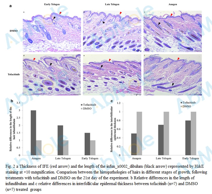 |
Source | Archives of Dermatological Research (2018). Figure 2. Tofacitinib (Abmole Bioscience, Houston, USA) |
| Method | i.v. | |
| Cell Lines | C57BL/6 mice | |
| Concentrations | concentration of 2% | |
| Incubation Time | 21 consecutive days | |
| Results | The length of the hair infundibulum on the 21st day of the experiment was higher in groups treated topically with tofacitinib. |
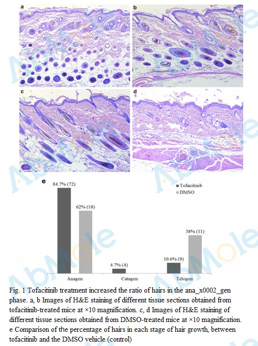 |
Source | Archives of Dermatological Research (2018). Figure 1. Tofacitinib (Abmole Bioscience, Houston, USA) |
| Method | i.v. | |
| Cell Lines | C57BL/6 mice | |
| Concentrations | concentration of 2% | |
| Incubation Time | 21 consecutive days | |
| Results | In mice treated topically with tofacitinib, 84.7% of hairs were in the anagen phase (n=72), 10.6% were in the catagen phase (n = 9), and 4.7% were in the telogen phase (n=4), where the follicles in the late anagen were located in the deep dermis and hypodermis, and follicles in the early anagen were located in the superficial dermis. |
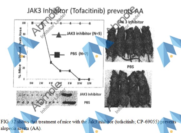 |
Source | US Patent 9730877B2 (2017). Figure 32. Tofacitinib (Abmole Bioscience) |
| Method | oral | |
| Cell Lines | mice | |
| Concentrations | 15 mg/kg/day | |
| Incubation Time | 12 weeks | |
| Results | None of the treated mice developed alopecia areata or cutaneous lymphadenopathy, whereas untreated mice manifested both AA and associated cutaneous lymphadenopathy |
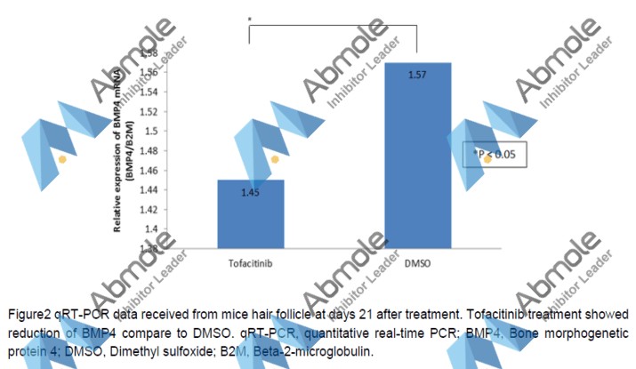 |
Source | TJPS (2017). Figure 2. Tofacitinib (Abmole Bioscience, Houston, TX, USA) |
| Method | qRT-PCR | |
| Cell Lines | mice hair follicle | |
| Concentrations | 2% concentration | |
| Incubation Time | 21 days | |
| Results | We investigate the expression of BMP4 which known to be an inhibitory molecule for onset of anagen entry. Quantitative real-time reverse transcription-PCR analysis showed decrease expression of BMP4 in tofacitinib treated group compare to DMSO-treated group (p-value 0.003). |
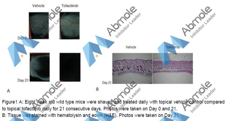 |
Source | TJPS (2017). Figure 1. Tofacitinib (Abmole Bioscience, Houston, TX, USA) |
| Method | Inject | |
| Cell Lines | C57BL/6 male mice | |
| Concentrations | 2% concentration | |
| Incubation Time | 21 days | |
| Results | We examined the effect of topical tofacitinib on hair growth in mice tissue, tofacitinib-treated group has shown denser in thickness and length of hair follicle compared to vehicle (Figure 1A). Histology, tofacitinib treated group has shown increased in anagen hair follicle compared to vehicle (Figure 1B). |
 |
Source | Arch Dermatol Res (2017). Figure 6. Tofacitinib (Abmole Bioscience, Texas, USA) |
| Method | HE stain | |
| Cell Lines | Mice | |
| Concentrations | dissolved in DMSO to create a 2% concentration | |
| Incubation Time | 21 day | |
| Results | Tofacitinib-treated mice showed less infiltration of inflammatory cells relative to minoxidil-treated mice. In the DMSO-treated group, infiltration of several inflammatory cells infiltration was observed, while fewer inflammatory cells were observed in the ethanol-treated group. |
 |
Source | Arch Dermatol Res (2017). Figure 5. Tofacitinib (Abmole Bioscience, Texas, USA) |
| Method | HE stain | |
| Cell Lines | Mice | |
| Concentrations | dissolved in DMSO to create a 2% concentration | |
| Incubation Time | 21 day | |
| Results | More newly formed capillaries were observed in the tofacitinib-treated group than in the minoxidil- and controltreated groups, although some completely formed capillaries were observed in the minoxidil- and DMSO-treated groups. |
 |
Source | Arch Dermatol Res (2017). Figure 4. Tofacitinib (Abmole Bioscience, Texas, USA) |
| Method | HE stain | |
| Cell Lines | Mice | |
| Concentrations | dissolved in DMSO to create a 2% concentration | |
| Incubation Time | 21 day | |
| Results | "At the end of the experiment, mostly anagen hairs were observed in the tofacitinib-treated group, whereas mostly catagen and anagen hairs were observed in minoxidil- and DMSO-treated groups." |
. ruxolitinib, figure 4..jpg) |
Source | Science Advance (2015). Ruxolitinib, Figure 4. (AbMole Bioscience) |
| Method | Patch assay with DP spheres | |
| Cell Lines | DP cells | |
| Concentrations | 400 nM | |
| Incubation Time | ||
| Results | In the region including genes thatwere upregulated by ruxolitinib treatment but down-regulated by tofacitinib treatment, we identified proapoptotic genes such as BAX, BCL2L11, and CASP12 (region 2). |
. ruxolitinib, figure 3..jpg) |
Source | Science Advance (2015). Ruxolitinib, Figure 3. (AbMole Bioscience) |
| Method | Human HF organ culture assay | |
| Cell Lines | human HFs | |
| Concentrations | 400 nM | |
| Incubation Time | ||
| Results | We found that treatment with JAK inhibitors significantly increased the length of hair shafts when treatedwith ruxolitinib and tofacitinib, indicating a positive effect on the rate of hair elongation (P = 0.023 and P = 0.025 for tofacitinib and ruxolitinib, respectively). |
. ruxolitinib, figure 1..jpg) |
Source | Science Advance (2015). Ruxolitinib, Figure 1. (AbMole Bioscience) |
| Method | Fourmice were treated with control (half of the back skin) and ruxolitinib (half of the back skin) and four mice were treated with control (half of the back skin) and tofacitinib (half of the back skin). | |
| Cell Lines | ||
| Concentrations | ||
| Incubation Time | ||
| Results | Indeed, ~90% of 8.5-week-old mice treated with ruxolitinib or tofacitinib for 5 days displayed skin darkening and hair growth within 10 days of starting treatment, whereas no hair growth was evident in control-treated mice (P < 0.0001 for ruxolitinib treatment and P = 0.04 for tofacitinib treatment). Ruxolitinib treatment enriched for the mTOR (mammalian target of rapamycin) and NFkB (nuclear factor kB) pathways, both previously shown to be involved in hair cycle regulation, whereas tofacitinib treatment enriched for pathways involved in cell motility and migration, such as Rho and integrin signaling. |
-2.jpg) |
Source | Nature medicine (2014). Figure 1. tofacitinib (Abmole) |
| Method | Flow cytometric analysis, Immunohistochemistry and immunofluorescence | |
| Cell Lines | ||
| Concentrations | 15 mg/kg/day | |
| Incubation Time | 12 weeks | |
| Results | "We observed complete hair regrowth within 12 weeks following topical therapy. Topical therapy was associated with a markedly reduced proportion of CD8+NKG2D+T cells in the treated skin and lymph node, normalization of the ALADIN transcriptional signature, reversal of histological markers of disease. " |
-1.jpg) |
Source | Nature medicine (2014). Figure 3. tofacitinib (Abmole) |
| Method | Flow cytometric analysis, Immunohistochemistry and immunofluorescence | |
| Cell Lines | ||
| Concentrations | 15 mg/kg/day | |
| Incubation Time | 12 weeks | |
| Results | we systemically administered tofacitinib at the time of grafting and found that they prevented the development of AA and the expansion of CD8+NKG2D+ T cells in all grafted recipients. The skin of mice treated with either drug showed no histological signs of inflammation. Global transcriptional analysis of whole-skin biopsies showed that both drugs also blocked the dermal inflammatory signature, as measured by Alopecia Areata Disease Activity Index. |
-2.jpg) |
Source | Nature medicine (2014). Figure 1. JAK3i tofacitinib (Abmole) |
| Method | grafted C3H/HeJ mice | |
| Cell Lines | CD8+NKG2D+ T cells | |
| Concentrations | 15 mg/kg/day | |
| Incubation Time | 12 weeks | |
| Results | we observed complete hair regrowth within 12 weeks following topical therapy. Topical therapy was associated with a markedly reduced proportion of CD8+NKG2D+ T cells in the treated skin and lymph node, normalization of the ALADIN transcriptional signature, reversal of histological markers of disease. |
-1.jpg) |
Source | Nature medicine (2014). Figure 1. JAK3i tofacitinib (Abmole) |
| Method | grafted C3H/HeJ mice | |
| Cell Lines | CD8+NKG2D+ T cells | |
| Concentrations | 15 mg/kg/day | |
| Incubation Time | 12 weeks | |
| Results | Global transcriptional analysis of whole-skin biopsies showed that both drugs also blocked the dermal inflammatory signature, as measured by Alopecia Areata Disease Activity Index |
| Cell Experiment | |
|---|---|
| Cell lines | cytokine-dependent NK92 cell line |
| Preparation method | (A) After cytokine starvation for 24 hours, NK92 cells were stimulated by the addition of human IL-2 for 48 hours with and without the addition of serially increasing concentrations of tofacitinib. 3H-thymidine was added during the last 6 hours of the cultures. Cells were then harvested and analyzed for 3H-thymidine incorporation. (B) Cytokine-starved NK92 cells were stimulated with human IL-2, combined IL-6/IL- 6R, or IL-12 for 48 hours with and without serially increasing concentrations of tofacitinib. (C) The NK92 cells were treated as those in panel B with and without a single 50nM dose of tofacitinib. Data are presented as means ±SD (A-C) and are representative of 3 independent experiments. (D) After cytokine starvation for 24 hours, NK92 cells were stimulated with 30 ng/mL of IL-2, 100 ng/mL of combined IL-6/IL-6R, or 100 ng/mL of IL-12 for 1 hour with and without the addition of tofacitinib. The cell lysates were immunoblotted with an anti–phospho-STAT5 monoclonal antibody and an anti-STAT5 antibody. ß-Actin was used as an input control. Data are representative of 3 independent experiments. |
| Concentrations | 0~400 nM |
| Incubation time | 48 h |
| Animal Experiment | |
|---|---|
| Animal models | IL-15–transgenic CD8 T-cell leukemia–bearing mice |
| Formulation | dissolved in polyethylene glycol 300 (PEG300; VWR Scientific Products) |
| Dosages | 50 mg/mL |
| Administration | continuously administered via a subcutaneous mini-osmotic pump (ALZET) |
| Molecular Weight | 312.37 |
| Formula | C16H20N6O |
| CAS Number | 477600-75-2 |
| Solubility (25°C) | DMSO ≥60 mg/mL |
| Storage | -20°C, sealed |
| Related JAK Products |
|---|
| iJak-381
iJak-381 is a JAK1/2 inhibitor with anti-inflammatory activity. |
| Ritlecitinib (malonate)
Ritlecitinib (PF-06651600) malonate is an orally active and selective JAK3 inhibitor with an IC50 of 33.1 nM. |
| Deuruxolitinib
Deuruxolitinib is a deuterated Ruxolitinib (INCB18424), which modulates the activity of JAK1/JAK2. Deuruxolitinib can be used for the research of hair loss disorders. |
| A-005
A-005 is a potentially first-in-class, brain-penetrating TYK2 allosteric inhibitor for studies related to neuroinflammatory and neurodegenerative diseases. |
| Tyrosine Protein Kinase JAK 2 (Phospho-Tyr8, 9)
Tyrosine Protein Kinase JAK 2 (Phospho-Tyr8, 9) is a peptide corresponding to amino acids 475 to 491 of mouse JAK2. |

All AbMole products are for research use only, cannot be used for human consumption or veterinary use. We do not provide products or services to individuals. Please comply with the intended use and do not use AbMole products for any other purpose.


Products are for research use only. Not for human use. We do not sell to patients.
© Copyright 2010-2024 AbMole BioScience. All Rights Reserved.