All AbMole products are for research use only, cannot be used for human consumption.
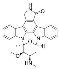
Staurosporine (antibiotic AM-2282 or STS) is a therapeutic agent that inhibits tumor cell growth by inducing cell death via intrinsic apoptotic pathways. Staurosporine is a prototypical ATP-competitive kinase inhibitor in that it binds to many kinases with high affinity, though with little selectivity. Staurosporine also inhibits a variety of other protein kinases, including PKA, PKG, phosphorylase kinase, S6 kinase, MLCK, CAM PKII, cdc2, v-Src, Lyn, c-Fgr and Syk with IC50 values of 15 nM, 18 nM, 3 nM, 5 nM, 21 nM, 20 nM, 9 nM, 6 nM, 20 nM, 2 nM and 16 nM respectively. Staurosporine induces apoptosis of human foreskin fibroblasts AG-1518, depending on the lysosomal cathepsins D mediated cytochrome c release and caspase activation. Moreover, Staurosporine triggers a novel intrinsic apoptosis pathway, relying on the activation of caspase-9 in the absence of Apaf-1.

Plant Physiology and Biochemistry. 2025 May 15; .
Mechanism of Silicon Nutrition Sensing in Salt-Stressed Rice Plants: Is It Modulated by FERONIA Homologs OsFLR1/2-Mediated Cell Wall Integrity Signaling?
Staurosporine purchased from AbMole
Inflamm Regen. 2023 Jun 20;43(1):31.
Promotion of axon regeneration and protection on injured retinal ganglion cells by rCXCL2
Staurosporine purchased from AbMole
Biomaterials. 2021 Aug;275:120958.
Tumor progress intercept by intervening in Caveolin-1 related intercellular communication via ROS-sensitive c-Myc targeting therapy
Staurosporine purchased from AbMole
Pak Vet J. 2021 Mar.
Evaluation of the PBMC Proliferation, Apoptosis and Cytokines Profiling in Cattle Infected with Mycoplasma bovis Strain 07801
Staurosporine purchased from AbMole
Patent. CN111089859A 2020 May 01.
Patent. CN111089859A
Staurosporine purchased from AbMole
PLoS One. 2019 Oct 10;14(10):e0223096.
Optimized bioluminescence analysis of adenosine triphosphate (ATP) released by platelets and its application in the high throughput screening of platelet inhibitors.
Staurosporine purchased from AbMole
Food Chem. 2017 Mar 15;219:304-310.
Effects of protein phosphorylation on color stability of ground meat
Staurosporine purchased from AbMole
Food Chem. 2017 Sep 20.
Dephosphorylation enhances postmortem degradation of myofibrillar proteins
Staurosporine purchased from AbMole
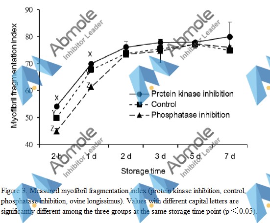 |
Source | Food Chem (2017). Figure 3. Staurosporine (Abmole Bioscience,Houston, TX, USA) |
| Method | Measured myofibril fragmentation index | |
| Cell Lines | Muscles | |
| Concentrations | 0.144 μmol/5 g | |
| Incubation Time | 2 h, 1 d, 2 d, 3 d, 5 d and 7 day | |
| Results | During the MFI increased phase (measured at 2 h and 1 d), the MFI values of the low phosphorylation level group (protein kinase inhibited group) were significantly higher (p < 0.05) than those of the control group, while the MFI values of the high phosphorylation level group (phosphatase inhibited group) were significantly lower (p < 0.05) than those of the control group. |
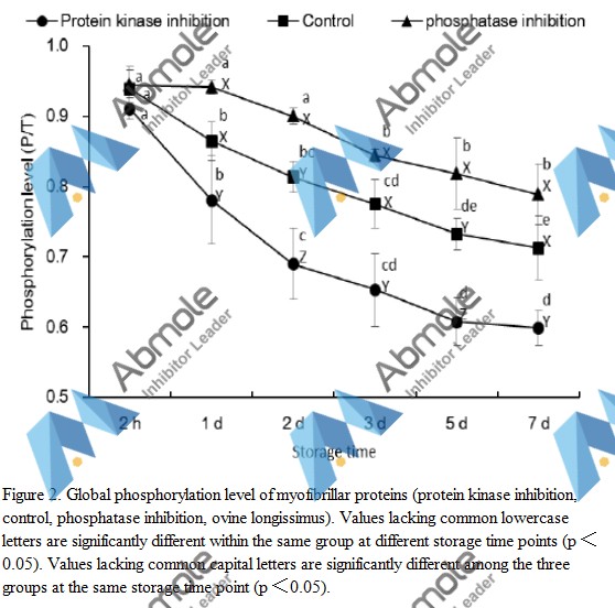 |
Source | Food Chem (2017). Figure 2. Staurosporine (Abmole Bioscience,Houston, TX, USA) |
| Method | Global phosphorylation level of myofibrillar proteins | |
| Cell Lines | Muscles | |
| Concentrations | 0.144 μmol/5 g | |
| Incubation Time | 2 h, 1 d, 2 d, 3 d, 5 d and 7 day | |
| Results | Inhibition of protein kinase by M2066 significantly reduced the phosphorylation level of myofibrillar proteins in muscle compared to control. |
.figure 4.jpg) |
Source | Food Chemistry (2016).Figure 4. kinase inhibitors (M2066, Abmole, Houston, TX, USA) |
| Method | Lactic acid concentrations | |
| Cell Lines | ||
| Concentrations | 0.144 μmole per 5 g muscle pieces | |
| Incubation Time | 2h, 24h, 48h, 72h, 120h and 168h | |
| Results | No difference was observed in lactic acid content (Fig. 4) between C1 and C2 control groups. In addition, no difference in lactic acid was detected between phosphatase inhibition group and the two control groups at most time points, but the kinase inhibition group had lower lactic acid content than the other three groups (P < 0.05) throughout the entire experimental period. |
.figure 3.jpg) |
Source | Food Chemistry (2016).Figure 3. kinase inhibitors (M2066, Abmole, Houston, TX, USA) |
| Method | PH Value | |
| Cell Lines | ||
| Concentrations | 0.144 μmole per 5 g muscle pieces | |
| Incubation Time | 2h, 24h, 48h, 72h, 120h and 168h | |
| Results | There was no difference in pH values (Fig. 3) between the phosphatase inhibition group and the two control groups. the pH of the kinase inhibition group declined much slower during the early postmortem stage (P < 0.05) as compared to the other three groups. |
.figure 2.jpg) |
Source | Food Chemistry (2016).Figure 2. kinase inhibitors (M2066, Abmole, Houston, TX, USA) |
| Method | protein phosphrylation assay | |
| Cell Lines | ||
| Concentrations | 0.144 μmole per 5 g muscle pieces | |
| Incubation Time | 2h, 24h, 48h, 72h, 120h and 168h | |
| Results | The R630/580 values (Fig. 2) were not different between C1 and C2 control groups at any time. However, the R630/580 values of the phosphatase inhibition group were lower than those of all other groups (P < 0.05) except at 2 h. Kinase inhibition group demonstrated greater (P < 0.05) R630/580 values than those of the rest groups except at 2 and 168 h. |
.figure 1.jpg) |
Source | Food Chemistry (2016).Figure 1. kinase inhibitors (M2066, Abmole, Houston, TX, USA) |
| Method | Gel electrophoresis and image analyses | |
| Cell Lines | ||
| Concentrations | 0.144 μmole per 5 g muscle pieces | |
| Incubation Time | 2h, 24h, 48h, 3d, 5d and 7d | |
| Results | "As expected, the overall phosphorylation of sarcoplasmic proteins in the minced meat with phosphatase inhibitor was significantly higher (P < 0.05) than in control and kinase inhibitor added samples." |
| Cell Experiment | |
|---|---|
| Cell lines | PC12 |
| Preparation method | Cells are exposed to Staurosporine for ~32 hours. Cells are fixed in 4% paraformaldehyde and stained with the DNA-binding dye Hoechst 33342. Cells are visualized under epifluorescence illumination, and the percentage of apoptotic cells (cells with condensed and fragmented DNA) is determined. |
| Concentrations | Dissolved in DMSO, final concentration 1 μM |
| Incubation time | ~32 hours |
| Animal Experiment | |
|---|---|
| Animal models | Male Mongolian gerbils or male Wistar rats subjected to transient ischemia |
| Formulation | Dissolved in DMSO, and diluted in saline |
| Dosages | ~10 ng |
| Administration | Stereotaxically administered into the bilateral CAl subfield of the hippocampus |
| Molecular Weight | 466.53 |
| Formula | C28H26N4O3 |
| CAS Number | 62996-74-1 |
| Solubility (25°C) | DMSO ≥ 50 mg/mL |
| Storage |
Powder -20°C 3 years ; 4°C 2 years In solvent -80°C 6 months ; -20°C 1 month |
| Related PKC Products |
|---|
| Sapintoxin D
Sapintoxin D is a fluorescent phorbol ester that can be used to study the activation of protein kinase C (PKC). |
| Bryostatin 3
Bryostatin 3, a macrocyclic lactone, is a protein kinase C activator, with a Ki of 2.75 nM. |
| Ruboxistaurin mesylate
Ruboxistaurin (LY333531) mesylate is an orally active, selective and ATP competitive PKCβ inhibitor with IC50 values of 4.7 and 5.9 nM for PKCβI and PKCβII, respectively. |
| PKC β pseudosubstrate
PKC β pseudosubstrate is a selective cell-permeable inhibitor of PKC. |
| Protein Kinase C (19-31)
Protein Kinase C (19-31), a peptide inhibitor of protein kinase C (PKC), is used as protein kinase C substrate peptide for testing the protein kinase C activity. |

All AbMole products are for research use only, cannot be used for human consumption or veterinary use. We do not provide products or services to individuals. Please comply with the intended use and do not use AbMole products for any other purpose.


Products are for research use only. Not for human use. We do not sell to patients.
© Copyright 2010-2024 AbMole BioScience. All Rights Reserved.