All AbMole products are for research use only, cannot be used for human consumption.

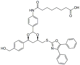
Niltubacin, as an inactive derivative of Tubacinis, is a highly potent and selective, reversible cell-permeable inhibitor of HDAC6. Histone deacetylase 6 (HDAC6) is structurally and functionally unique among the 11 human zinc-dependent histone deacetylases. HDAC6-selective inhibitor tubacin significantly enhances cell death induced by the topoisomerase II inhibitors etoposide and doxorubicin and the pan-HDAC inhibitor SAHA (vorinostat) in transformed cells (LNCaP, MCF-7), an effect not observed in normal cells. The inactive analogue of tubacin, nil-tubacin, does not sensitize transformed cells to these anticancer agents.

EMBO J. 2019 Jan 15;38(2).
Prominin-1 controls stem cell activation by orchestrating ciliary dynamics.
Niltubacin purchased from AbMole

J Cell Biochem. 2017 Apr 27.
The HDAC6 Inhibitor Tubacin Induces Release of CD133þ Extracellular Vesicles From Cancer Cells
Niltubacin purchased from AbMole

Mol Cell Biol. 2016 Oct 28;36(22):2838-2854.
Activated Transcription Factor 3 in Association with Histone Deacetylase 6 Negatively Regulates MicroRNA 199a2 Transcription by Chromatin Remodeling and Reduces Endothelin-1 Expression.
Niltubacin purchased from AbMole
| Cell Experiment | |
|---|---|
| Cell lines | Raji cells |
| Preparation method | Cell adhesion assay Prior to SDF-1α (100 ng/ml) stimulation, Raji cells were treated with DMSO, NaB (1 μM), or tubacin (1 μM) for 3 hours. Then cells were plated into a 96-well plate which were precoated with Fibronectin (50 ng/ml) to allow cell adherence for 30 minutes. Subsequently cells were washed with PBS three times to remove the non-adherent cells, and adherent cells were measured by CellTiter-Glo luminescent cell viability assay kit. |
| Concentrations | 1μM |
| Incubation time | 3h |
| Animal Experiment | |
|---|---|
| Animal models | PLD animal model 3-week-old PCK rats |
| Formulation | saline |
| Dosages | 30 mg/kg body |
| Administration | intraperitoneally |
| Molecular Weight | 706.85 |
| Formula | C41H42N2O7S |
| Solubility (25°C) | DMSO ≥5 mg/mL |
| Storage |
Powder -20°C 3 years ; 4°C 2 years In solvent -80°C 6 months ; -20°C 1 month |
| Related HDAC Products |
|---|
| CM-444
CM-444 is an inhibitor for HDAC and DNA methyltransferases (DNMT) with IC50 values of 6 nM-0.6 μM and 1.8-2.3 μM, respectively. CM-444 is an inducer for the differentiation of acute myeloid leukemia cells. CM-444 exhibits anti-leukemic activity and improves the survival rate in mouse models. |
| CM-1758
CM-1758 is a histone deacetylase (HDAC) inhibitor. CM-1758 inhibits tumor growth in vivo. CM-1758 induces acetylation of non-histone proteins in acute myeloid leukemia cells. |
| T-518
T-518 is an orally active, selective, and blood-brain barrier permeable HDAC6 inhibitor with an IC50 value of 36 nM for human HDAC6. |
| SE-7552
SE-7552, a 2-(difluoromethyl)-1,3,4-oxadiazole (DFMO) derivative, is an orally active, highly selective, non-hydroxamate HDAC6 inhibitor with an IC50 of 33 nM. |
| BRD9757
BRD9757 is a potent, capless and selective HDAC6 inhibitor with an IC50 of 30 nM. |
All AbMole products are for research use only, cannot be used for human consumption or veterinary use. We do not provide products or services to individuals. Please comply with the intended use and do not use AbMole products for any other purpose.


Products are for research use only. Not for human use. We do not sell to patients.
© Copyright 2010-2024 AbMole BioScience. All Rights Reserved.
