All AbMole products are for research use only, cannot be used for human consumption.
GDC-0941, a selective and potent PI3K inhibitor, is highly efficacious both in combination with trastuzumab and in the treatment of trastuzumab-resistant cells and tumors. GDC-0941 inhibited the growth of >70% of breast cancer cell lines tested, with EC50's <1 µM. GDC-0941 also demonstrated antitumor activity in preclinical models of breast cancer.

Patent. WO2020115009A1 2020 Jun 11.
Patent. WO2020115009A1
GDC-0941 (Pictilisib) purchased from AbMole
J Nucl Med. 2018 Nov 21.
MEK inhibition induces therapeutic iodine uptake in a murine model of anaplastic thyroid cancer.
GDC-0941 (Pictilisib) purchased from AbMole

J Exp Clin Cancer Res. 2018 Sep 21;37(1):234.
The LAT1 inhibitor JPH203 reduces growth of thyroid carcinoma in a fully immunocompetent mouse model.
GDC-0941 (Pictilisib) purchased from AbMole
Mol Cancer Res. 2018 Aug 14.
Comprehensive Genomic Profiling of Patient-matched Head and Neck Cancer Cells: A Preclinical Pipeline for Metastatic and Recurrent Disease.
GDC-0941 (Pictilisib) purchased from AbMole
ChemMedChem. 2018 Dec 6.
Identifying Lysophosphatidic Acid Acyltransferase β (LPAAT-β) as the Target of a Nanomolar Angiogenesis Inhibitor from a Phenotypic Screen Using the Polypharmacology Browser PPB2.
GDC-0941 (Pictilisib) purchased from AbMole

Mol Cancer. 2017 May 22;16(1):93.
PIK3CA hotspot mutations differentially impact responses to MET targeting in MET-driven and non-driven preclinical cancer models
GDC-0941 (Pictilisib) purchased from AbMole
Oncotarget. 2017 Apr 11;8(15): 24604–24620.
Combined MEK and Pi3’-kinase inhibition reveals synergy in targeting thyroid cancer in vitro and in vivo
GDC-0941 (Pictilisib) purchased from AbMole
BioRxiv. 2017 July 18;1-45.
PIK3CAH1047R-induced paradoxical ERK activation results in resistance to BRAFV600E specific inhibitors in BRAFV600E PIK3CAH1047R double mutant thyroid tumors.
GDC-0941 (Pictilisib) purchased from AbMole
| Source | Journal of Nuclear Medicine (2018 Nov). Figure 3. GDC-0941 (AbMole Bioscience Inc.) | |
| Method | oral gavage | |
| Cell Lines | single mutant BRAFV600E mice | |
| Concentrations | 50 mg/kg | |
| Incubation Time | 10 days | |
| Results | The selective BRAFV600E inhibitor PLX-4720 did not increase Nis mRNA transcription, nor did the PI3K inhibitor GDC-0941 alone or in combination with PD-325901 or PLX-4720. |
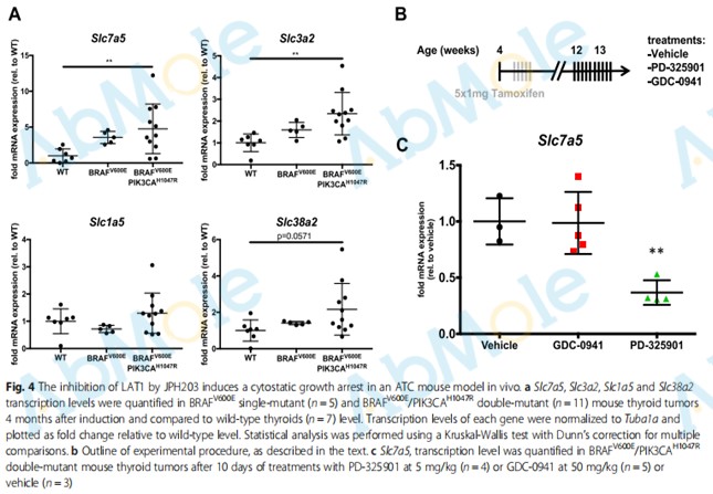 |
Source | Journal of Experimental & Clinical Cancer Research (2018). Figure 4. GDC-0941 (Abmole Bioscience) |
| Method | oral gavage | |
| Cell Lines | BRAFV600E/PIK3CAH1047R double-mutant mouse | |
| Concentrations | 50 mg/kg | |
| Incubation Time | 10 days | |
| Results | While Pi3K inhibition did not induced any change in Slc7a5 transcript abundance, MEK inhibition induced more than 50% reduction. |
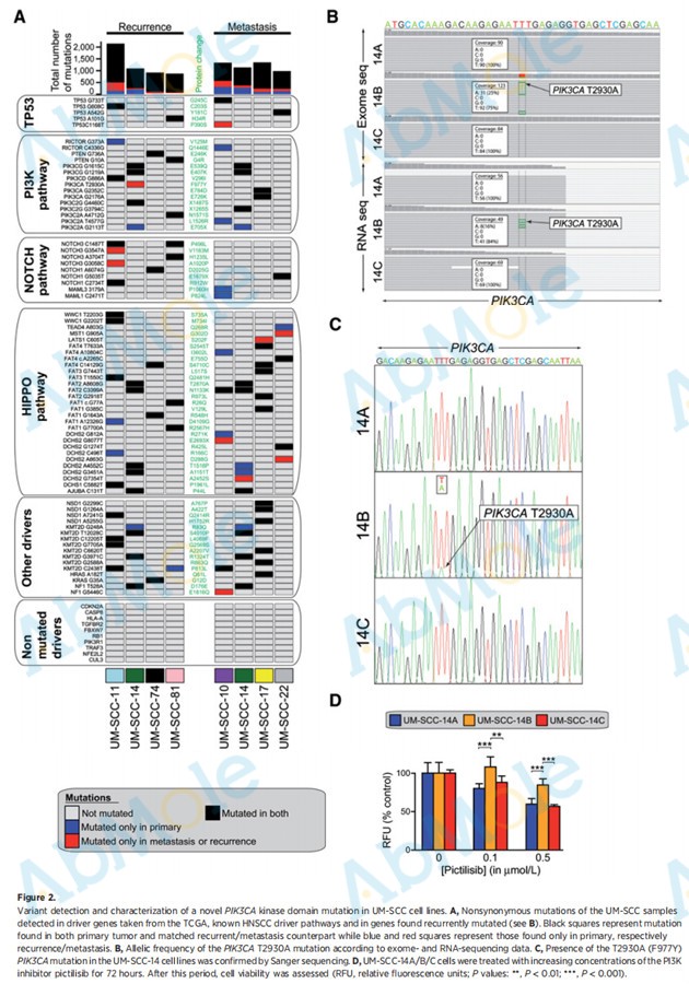 |
Source | Molecular Cancer Research (2018). Figure 2. GDC-0941 (Abmole Bioscience) |
| Method | cell viability assay | |
| Cell Lines | M-SCC-14A, UM-SCC-14B, and UM-SCC-14C cells | |
| Concentrations | ||
| Incubation Time | 72 hours | |
| Results | Interestingly, pictilisib was less effective at impairing proliferation in UM-SCC-14B than in the other two matched cell lines. |
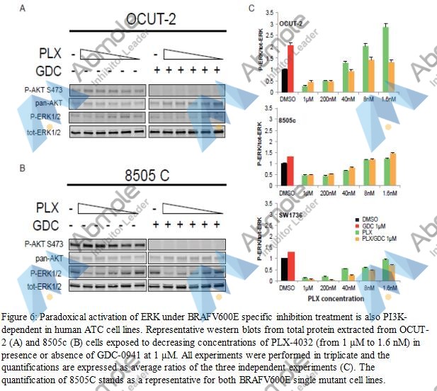 |
Source | BioRxiv (2017). Figure 6. GDC-0941 (Abmole Bioscience, Hong-Kong, China) |
| Method | western blot | |
| Cell Lines | 8505c cells | |
| Concentrations | 1 μM | |
| Incubation Time | ||
| Results | Paradoxical ERK hyperphosphorylation was not detectable when cells were subjected to GDC-0941 in addition to PLX-4032 drug dilutions. When we treated them with the same concentrations of PLX-4032, 8505c and SW1736 cells did not exhibit paradoxical ERK activation. |
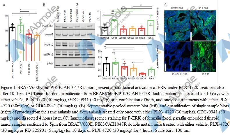 |
Source | BioRxiv (2017). Figure 4. GDC-0941 (Abmole Bioscience, Hong-Kong, China) |
| Method | paradoxical activation | |
| Cell Lines | Thyroglobulin Cre ERT2 mice | |
| Concentrations | 50 mg/kg | |
| Incubation Time | 10 d | |
| Results | Only GDC-0941 and drug combination treated animals showed tumor burden reduction. PLX-4720 treated animals displayed an elevation in tumor burden that was not statistically different from the controls but from the two other groups. |
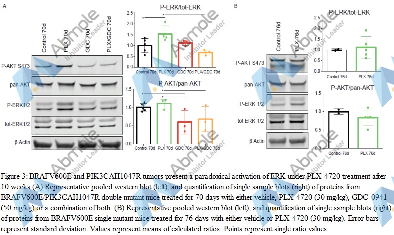 |
Source | BioRxiv (2017). Figure 3. GDC-0941 (Abmole Bioscience, Hong-Kong, China) |
| Method | paradoxical activation | |
| Cell Lines | Thyroglobulin Cre ERT2 mice | |
| Concentrations | 50 mg/kg | |
| Incubation Time | 70 d | |
| Results | When treated with drug combination, ERK paradoxical activation was abolished resulting in ERK phosphorylation level comparable to controls. AKT phosphorylation was not affected by PLX-4720 while GDC-0941 treatment resulted in a small but significant reduction of AKT phosphorylation. |
 |
Source | BioRxiv (2017). Figure 2. GDC-0941 (Abmole Bioscience, Hong-Kong, China) |
| Method | Histological presentation | |
| Cell Lines | Thyroglobulin Cre ERT2 mice | |
| Concentrations | 50 mg/kg | |
| Incubation Time | 70 d | |
| Results | GDC-0941-treated mice had smaller thyroid sections, while presenting a similar histology compared to controls (Fig. 2B) with a mixture of PTC containing small ATC foci. |
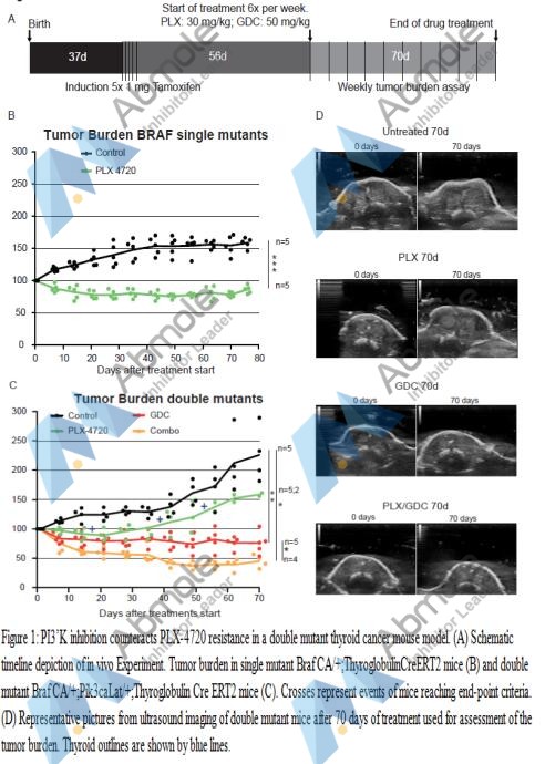 |
Source | BioRxiv (2017). Figure 1. GDC-0941 (Abmole Bioscience, Hong-Kong, China) |
| Method | tumor burden assay | |
| Cell Lines | Thyroglobulin Cre ERT2 mice | |
| Concentrations | 50 mg/kg | |
| Incubation Time | 70 d | |
| Results | GDC-0941 treated animals presented an initial tumor burden reduction of 20% then tumor size stabilized for the rest of the treatment period. Interestingly, when a drug combination of PLX-4720 and GDC-0941 was administered, mice showed a robust response with 60% lower tumor burden after 6 weeks followed by stabilization until the end of the experiment. |
.figure 4..jpg) |
Source | Oncotarget (2017). GDC-0941, Figure 4. (AbMole Bioscience, Hong-Kong, China) |
| Method | Western blot | |
| Cell Lines | ATC cell | |
| Concentrations | 50 mg/kg | |
| Incubation Time | 24 h | |
| Results | Interestingly, PD-325901 treated mice showed a clear improvement in histology with some almost normal follicles and smaller PTC areas. GDC-0941 did not induce a beneficial effect at the histological level. Finally, mice treated with the combination, although resulting in smaller sections, seemed to have a similar histological presentation to PD-325901 alone treated animals (Figure 4C). |
.figure 3..jpg) |
Source | Oncotarget (2017). GDC-0941, Figure 3. (AbMole Bioscience, Hong-Kong, China) |
| Method | Western blot | |
| Cell Lines | ATC cell | |
| Concentrations | ||
| Incubation Time | 24 h | |
| Results | ERK1/2 and AKT phosphorylation were assessed first to demonstrate the drug efficiency. ERK1/2 phosphorylation ratio (p-ERK1/2 normalized to total ERK) was strongly decreased in all cell lines when treated with PD-325901 alone or in combination with GDC-0941. Similarly, GDC-0941 induced a strong reduction of AKT phosphorylation ratio (Figure 3). |
.figure 2..jpg) |
Source | Oncotarget (2017). GDC-0941, Figure 2. (AbMole Bioscience, Hong-Kong, China) |
| Method | apoptosis assay | |
| Cell Lines | OCUT-2 cells | |
| Concentrations | 1 μM | |
| Incubation Time | 24 h | |
| Results | Only the OCUT-2 cell line already showed increased apoptosis (double positive annexinV and PI cells) when treated with the combination for 24 h (Figure 2A). However, after 48 h of combination treatment, all three cell lines (Figure 2B and Supplementary Figure 1) had elevated double positive annexinV/PI cells (late apoptosis) and annexinV positive cells (early apoptosis). |
.figure 1..jpg) |
Source | Oncotarget (2017). GDC-0941, Figure 1. (AbMole Bioscience, Hong-Kong, China) |
| Method | ||
| Cell Lines | SW1736 and OCUT-2 cell lines | |
| Concentrations | 2 μM, 400 nM, 80 nM, 16 nM, 3.2 nM | |
| Incubation Time | 72 h | |
| Results | We investigated the effect of the drugs on cell cycling. PD-325901 alone or in combination with GDC-0941 induced a G1 cycle arrest in SW1736 and 8505c cell lines. However, in OCUT-2, a significant effect was only observed for the combination (Figure 1C). |
. gdc-0941, figure 5.jpg) |
Source | Mol Cancer (2017). GDC-0941, Figure 5. (AbMole BioScience, Hongkong, China) |
| Method | cell proliferation assay | |
| Cell Lines | NIH3T3 cells | |
| Concentrations | 0-100 nM | |
| Incubation Time | 16 h | |
| Results | Combination treatment led to more effective abrogation of AKT phosphorylation than either tepotinib or pictilisib alone, but p-S6 levels were generally unaltered (Fig. 5b). |
. gdc-0941, figure 4.jpg) |
Source | Mol Cancer (2017). GDC-0941, Figure 4. (AbMole BioScience, Hongkong, China) |
| Method | In vivo tumor growth delay experiments | |
| Cell Lines | ||
| Concentrations | 50 mg/kg | |
| Incubation Time | 5 d | |
| Results | Comparison of average tumor sizes of vector vs. H1047R at the experimental endpoint showed significant resistance to tepotinib in H1047R tumors (p = 0.012), as well as higher efficacy of pictilisib in the same group (p = 0.026; Fig. 4d). |
. gdc-0941, figure 1.jpg) |
Source | Mol Cancer (2017). GDC-0941, Figure 1. (AbMole BioScience, Hongkong, China) |
| Method | Cell viability/toxicity assays | |
| Cell Lines | NIH3T3 cells | |
| Concentrations | 0-100 nM | |
| Incubation Time | 16 h | |
| Results | PI3K inhibition by pictilisib was similarly effective in reducing p-AKT levels in these three cell lines, but at a concentration of 100 nM p-S6 levels were lower in cells harboring PIK3CA mutations (Fig. 1b). |
| Cell Experiment | |
|---|---|
| Cell lines | U87MG cell line |
| Preparation method | Proliferation Assay. The human tumor cell lines used wereobtained from the ATCC. Cells were plated at 4 × 10⁴ cells/ mL and cultured at 37 °C with 5% CO2 in DMEM supplemented with 10% fetal calf serum, and L-glutamine. Test compound was added to replicate wells in a volume of 10 µL such that the final DMSO concentration did not exceed 0.2%. After 4 days of incubation, 10 µL of Alamar Blue reagent was added and developed for 6 h at 37 °C before measuring the fluorescence excitation/emission (wavelength 540/595 nm) using a Victor plate reader. The reported IC50 values are means of at least two independent experiments with variations of less than 20%. |
| Concentrations | 0~10µM |
| Incubation time | 4 days |
| Animal Experiment | |
|---|---|
| Animal models | Human tumor xenografts of U87MG glioblastoma of female NCr athymic mice |
| Formulation | 10% DMSO, 5% Tween 20, 85% water |
| Dosages | 75 mg/kg once daily |
| Administration | oral gavage |
| Molecular Weight | 513.64 |
| Formula | C23H27N7O3S2 |
| CAS Number | 957054-30-7 |
| Solubility (25°C) | DMSO 40 mg/mL |
| Storage |
Powder -20°C 3 years ; 4°C 2 years In solvent -80°C 6 months ; -20°C 1 month |
| Related PI3K Products |
|---|
| T-00127-HEV1
T-00127-HEV1 is a phosphatidylinositol 4-kinase III beta (PI4KB) inhibitor with an IC50 of 60 nM. |
| PI3Kγ inhibitor AZ2
PI3Kγ inhibitor AZ2 is a highly selective PI3Kγ inhibitor (The pIC50 value for PI3Kγ is 9.3). |
| NIBR-17
NIBR-17 is a pan-class I PI3K inhibitor with suitable pharmacokinetic properties and inhibits tumor growth. |
| RV-1729
RV-1729 is an inhibitor of the phosphatidylinositol 3-kinase-δ (PI3Kδ). |
| Vulolisib
Vulolisib is a potent and orally active phosphatidylinositol 3-kinase (PI3K) inhibitor, with IC50 values of 0.2 nM, 168 nM, 90 nM and 49 nM for PI3Kα, PI3Kβ, PI3Kγ and PI3Kδ, respectively. |

All AbMole products are for research use only, cannot be used for human consumption or veterinary use. We do not provide products or services to individuals. Please comply with the intended use and do not use AbMole products for any other purpose.


Products are for research use only. Not for human use. We do not sell to patients.
© Copyright 2010-2024 AbMole BioScience. All Rights Reserved.