All AbMole products are for research use only, cannot be used for human consumption.
DAPT (GSI-IX) is a γ-secretase inhibitor with IC50 values of 115 and 200 nM for total Aβ and Aβ42 respectively. DAPT (GSI-IX) may be useful in the study of β-amyloid (Aβ) formation. As an inhibitor of Notch, DAPT may advance studies of autoimmune and lymphoproliferative diseases, such as ALPS and lupus erythematosus (SLE). Other γ-secretase substrates include LDL receptor-related protein, E-cadherin and ErbB-4.

J Obstet Gynaecol Res. 2025 May 01;51(5):e16295.
NINJ1 exerts a role in the development of preeclampsia through potential regulation by the Notch1 signaling pathway
DAPT purchased from AbMole
Front Surg. 2023 Jan 16;9:1027067.
Bacterial cellulose membrane combined with BMSCs promotes wound healing by activating the notch signaling pathway
DAPT purchased from AbMole
Glycoconj J. 2022 Jun 6.
Notch/NICD/RBP-J signaling axis regulates M1 polarization of macrophages mediated by advanced glycation end products
DAPT purchased from AbMole
J Ethnopharmacol. 2019 Oct 28;243:112092.
Study of the aqueous extract of Aloe vera and its two active components on the Wnt/β-catenin and Notch signaling pathways in colorectal cancer cells.
DAPT purchased from AbMole

Circ Res. 2017 Jul 31.
SUMOylation Negatively Regulates Angiogenesis by Targeting Endothelial NOTCH Signaling
DAPT purchased from AbMole

Oncol Lett. 2017 July 5;4: 3616-3622.
The role of Jagged1/Notch pathway‑mediated angiogenesis of hepatocarcinoma cells in vitro, and the effects of the spleen‑invigorating and blood stasis‑removing recipe
DAPT purchased from AbMole

Sci Adv. 2015 Apr 10.
PlGF-induced VEGFR1-dependent vascular remodeling determines opposing antitumor effects and drug resistance to Dll4-Notch inhibitors.
DAPT purchased from AbMole
2015 May.
Acquisition of resistance to trastuzumab in gastric cancer cells is associated with activation of IL-6/STAT3/Jagged-1/Notch positive feedback loop
DAPT purchased from AbMole
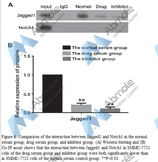 |
Source | Oncol Lett (2017). Figure 6. DAPT (AbMole BioScience, Shanghai, China) |
| Method | Co‑IP assay | |
| Cell Lines | SMMC‑7721 cells | |
| Concentrations | 1 μM | |
| Incubation Time | ||
| Results | The interaction between Jagged1 and Notch1 in SMMC‑7721 cells was also significantly reduced after adding DAPT compared with that of the normal serum group (P<0.01) (Fig. 6). |
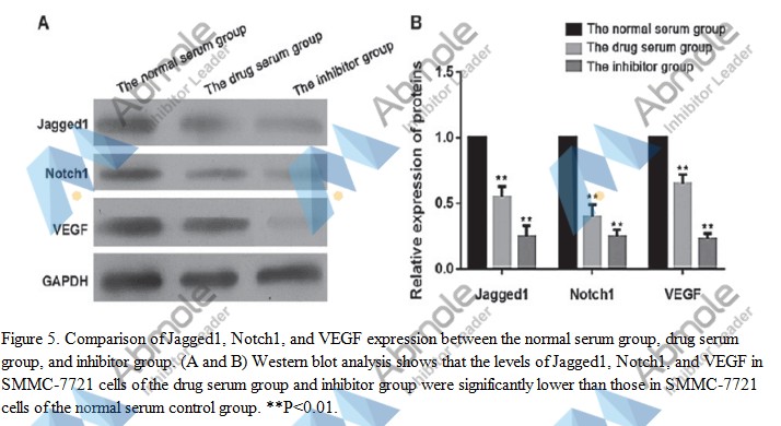 |
Source | Oncol Lett (2017). Figure 5. DAPT (AbMole BioScience, Shanghai, China) |
| Method | Western blot | |
| Cell Lines | SMMC‑7721 cells | |
| Concentrations | 1 μM | |
| Incubation Time | ||
| Results | The levels of Jagged1 and Notch1 were decreased after adding the Notch inhibitor, DAPT, indicating that DAPT successfully inhibited the Jagged1/Notch signaling pathway. In addition, the expression of VEGF protein was reduced after adding DAPT (Fig. 5). |
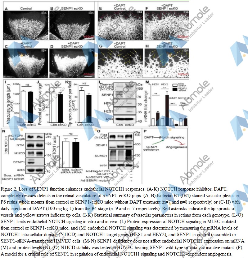 |
Source | Circ Res (2017). Figure 2. DATP (Abmole Bioscience) |
| Method | Retina dissection and whole mount staining | |
| Cell Lines | mice | |
| Concentrations | 100 mg/kg | |
| Incubation Time | ||
| Results | DAPT treatment resulted in a large increase in vessel density and expansion of the vascular plexus in the postnatal retina of control mice (Fig. 2A, C, E, G). DAPT completely rescued the defect in SENP1-ecKO pups (Fig. 2B, D, F, H), which resulted in vascular length, vascular coverage, and tip sprouts growth comparable to the control group (Fig. 2I-K). |
-4.jpg) |
Source | Sci adv (2015). Figure 5. The DAPT g-secretase inhibitor was obtained from AbMole BioScience. |
| Method | Immunohistochemistry | |
| Cell Lines | ||
| Concentrations | 100 mg/kg, daily | |
| Incubation Time | ||
| Results | "The PlGF-modulated opposing vascular effects in relation to Dll4-Notch inhibition alter tumor growth by changing the microenvironment, tumor cell proliferation, and apoptosis." |
-3.jpg) |
Source | Sci adv (2015). Figure 4. The DAPT g-secretase inhibitor was obtained from AbMole BioScience. |
| Method | human tumor models, Immunohistochemistry | |
| Cell Lines | ||
| Concentrations | 100 mg/kg, daily | |
| Incubation Time | ||
| Results | PlGF mediates opposing vascular effects in nontreated and Dll4-Notch inhibitor–treated mouse tumors. |
-2.jpg) |
Source | Sci adv (2015). Figure 3. The DAPT g-secretase inhibitor was obtained from AbMole BioScience. |
| Method | Immunohistochemistry | |
| Cell Lines | Human JE-3, BeWo, MDA-MB-231 cells | |
| Concentrations | 100 mg/kg, daily | |
| Incubation Time | ||
| Results | "Both terminal deoxynucleotidyl transferase–mediated deoxyuridine triphosphate nick end labeling (TUNEL) staining and measurements of the activated or cleaved caspase 3 showed reductions of apoptotic cells in Dll4-Notch inhibitor–treated JE-3 and BeWo tumors. In contrast, treatment of PlGF-negative MDAMB-231 tumors with DAPT and Dll4 blockade showed completely opposite effects on cell proliferation and apoptosis, tipping the balance toward an apoptotic phenotype." |
-1.jpg) |
Source | Sci adv (2015). Figure 1. The DAPT g-secretase inhibitor was obtained from AbMole BioScience. |
| Method | Immunohistochemistry | |
| Cell Lines | Human JE-3, BeWo, MDA-MB-231 cells | |
| Concentrations | 100 mg/kg, daily | |
| Incubation Time | ||
| Results | "PlGF expression determines opposing effects of Dll4-Notch inhibition on human tumor growth and vascular remodeling." |
| Cell Experiment | |
|---|---|
| Cell lines | SK-MES-1 |
| Preparation method | Cells are seeded into 96-well plates and exposed to 0.1% DMSO or DAPT at concentrations in the range of 2.5 μM–160 μM for 72 hours. Cytotoxicity is determined with 3-(4, 5)-dimethylthiahiazo-(-z-y1)-3, 5-di-phenytetrazoliumromide (MTT) dye reduction assay with minor modifications. Briefly, after incubation with DAPT, 20 μL MTT solution (5 mg/mL in PBS) is added to 180 μL medium in each well and plates are incubated for 4 hours at 37 °C, and subsequently 150 μL DMSO is added to each well, and mixed by shaking at room temperature for 15 minutes. Absorption is measured by an enzyme-linked immunosorbent assay at 490 nm to determine absorbance values. |
| Concentrations | 2.5 ~ 160 μM |
| Incubation time | 72 h |
| Animal Experiment | |
|---|---|
| Animal models | three‐ to four‐month‐old heterozygous PDAPP transgenic mice overexpressing the APPV717F mutant form of the amyloid precursor protein |
| Formulation | formulated in corn oil, 5% (v/v) ethanol |
| Dosages | 100 mg/kg |
| Administration | s.c. |
| Molecular Weight | 432.46 |
| Formula | C23H26F2N2O4 |
| CAS Number | 208255-80-5 |
| Solubility (25°C) | DMSO 60 mg/mL |
| Storage |
Powder -20°C 3 years ; 4°C 2 years In solvent -80°C 6 months ; -20°C 1 month |
| Related Gamma-secretase/Beta-secretase Products |
|---|
| Nirogacestat dihydrobromide
Nirogacestat dihydrobromide (PF-3084014 dihydrobromide) is a reversible, orally bioavailable, noncompetitive, and selective γ-secretase inhibitor with an IC50 of 6.2 nM. |
| RE(EDANS)EVNLDAEFK(DABCYL)R
RE (EDANS) EVNLDAEFK (DABCYL) R is an EDANS and DABCYL double-labeled peptide,serves as a fluorescent substrate for BACE1(Em=360nm,Ex=528nm). |
| β-Secretase inhibitor
β-Secretase inhibitor ([Asn670, Sta671, Val672]-Amyloid β Peptide (662-675)) is a β-secretase and BACE1 inhibitor (IC50: 25 nM for β-secretase). |
| Mca-SEVNLDAEFK(Dnp)
Mca-SEVNLDAEFK(Dnp) is a Beta-secretase 1 (BACE-1) peptide FRET substrate, containing the 'Swedish' Lys-Met/Asn-Leu mutation of the amyloid precursor protein (APP) β-secretase cleavage site. |
| BACE1-IN-10
BACE1-IN-10 is a potent BACE1 Inhibitor. |

All AbMole products are for research use only, cannot be used for human consumption or veterinary use. We do not provide products or services to individuals. Please comply with the intended use and do not use AbMole products for any other purpose.


Products are for research use only. Not for human use. We do not sell to patients.
© Copyright 2010-2024 AbMole BioScience. All Rights Reserved.