ABT-737 is a potent and selective inhibitor of B-cell lymphoma-2 (BCL-2) family proteins. The ABT-737 single-agent LC90 values ranged from 100 nM for COG-LL-319 and RS4; 11 cells to low micromolar concentrations for the other five ALL cell lines. ABT-737 efficiently induces apoptosis and suppresses growth of hepatoma cells in combination with sorafenib. Moreover, we observed that the enhancement of ABT-737-mediated apoptosis by NCTD was associated with activation of mitochondrial apoptosis signaling pathway, which involved cytosolic release of cytochrome c, cleavage of caspase-9 and caspase-3. Finally, knockdown of Mcl-1 substantially potentiated ABT-737-mediated apoptotic cell death, confirming the potency of Mcl-1 repression by NCTD in enhancing ABT-737-induced apoptosis. These results therefore suggest that combination treatment with NCTD can overcome ABT-737 resistance and enhance ABT-737 therapeutic efficacy in treating human HCC.
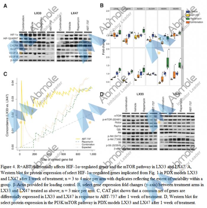 |
Source | Cancer Res (2014). Figure 4. ABT-737 |
| Method | Western blot | |
| Cell Lines | SCLC cell lines | |
| Concentrations | 100 nmol/L | |
| Incubation Time | 24 or 72 h | |
| Results | We did observe that the proapoptotic proteins BAX and BAK were increased in both PDXs treated with rapamycin alone or in combination with ABT-737. |
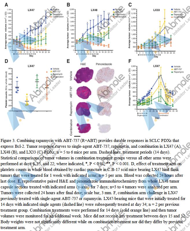 |
Source | Cancer Res (2014). Figure 3. ABT-737 |
| Method | i.e. | |
| Cell Lines | SCLC PDX models | |
| Concentrations | 100 mg/kg | |
| Incubation Time | 14 d | |
| Results | Correlating these findings with transcriptional profiling data, we observed decreased levels of six HIF-1α–regulated genes in LX47 after treatment with ABT-737, this effect was less pronounced in LX33, but nonetheless, observed for most genes. |
| Cell Experiment | |
|---|---|
| Cell lines | CLL cells and platelets |
| Preparation method | Apoptosis analysis Peripheral blood mononuclear cells were purified using Histopaque and cultured in RPMI 1640 medium supplemented with 10% fetal calf serum and 2 mmol/l L-glutamine at2*106 cells/ml. Cells were exposed to different concentrations of ABT-737, ABT-263, or ABT-199 for 4 h before analysis of apoptosis by externalisation of phosphatidylserine (PS) and binding of AnnexinV/fluorescein isothiocyanate (FITC). Platelets isolated from healthy volunteers were cultured in Hepes-buffered saline and exposed to different concentrations of ABT-737, ABT-263, or ABT-199 for 4 h before analysis of apoptosis in the CD41-positive population by externalisation of PS and binding of AnnexinV/FITC. |
| Concentrations | 0~3µM |
| Incubation time | 4 h |
| Animal Experiment | |
|---|---|
| Animal models | C57BL/6 mice were injected (i.v.) with 3x10⁶ T1 lymphoma cells |
| Formulation | formulated in 60% phosal 50PG (standardized phosphatidylcholine concentrate with at least 50% PC and propylene glycol |
| Dosages | 100 mg/kg |
| Administration | oral gavage |
| Molecular Weight | 813.43 |
| Formula | C42H45ClN6O5S2 |
| CAS Number | 852808-04-9 |
| Solubility (25°C) | DMSO 60 mg/mL |
| Storage |
Powder -20°C 3 years ; 4°C 2 years In solvent -80°C 6 months ; -20°C 1 month |
| Species | Mouse | Rat | Rabbit | Guinea pig | Hamster | Dog |
| Weight (kg) | 0.02 | 0.15 | 1.8 | 0.4 | 0.08 | 10 |
| Body Surface Area (m2) | 0.007 | 0.025 | 0.15 | 0.05 | 0.02 | 0.5 |
| Km factor | 3 | 6 | 12 | 8 | 5 | 20 |
| Animal A (mg/kg) = Animal B (mg/kg) multiplied by | Animal B Km |
| Animal A Km |
For example, to modify the dose of Compound A used for a mouse (20 mg/kg) to a dose based on the BSA for a rat, multiply 20 mg/kg by the Km factor for a mouse and then divide by the Km factor for a rat. This calculation results in a rat equivalent dose for Compound A of 10 mg/kg.
| Related Bcl-2 Products |
|---|
| BAD (103-127) (human)
BAD (103-127) (human), the 25-mer Bad peptide, is derived from the BH3 domain of BAD, can antagonize the function of Bcl-xL. |
| Bim BH3, Peptide IV
Bim BH3, Peptide IV is a 26-residue peptide from BH3-only protein Bim, which belongs to the pro-apoptotic group of the Bcl-2 family of proteins. |
| Bax inhibitor peptide, negative control
Bax inhibitor peptide, negative control is a inhibitor of Bax. |
| Bad BH3 (mouse)
Bad BH3 (mouse) is a biological active peptide. |
| Bid BH3 (80-99)
Bid BH3 (80-99) is a biological active peptide. |


Products are for research use only. Not for human use. We do not sell to patients.
© Copyright 2010-2023 AbMole BioScience. All Rights Reserved.